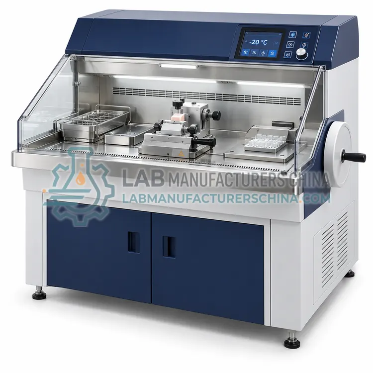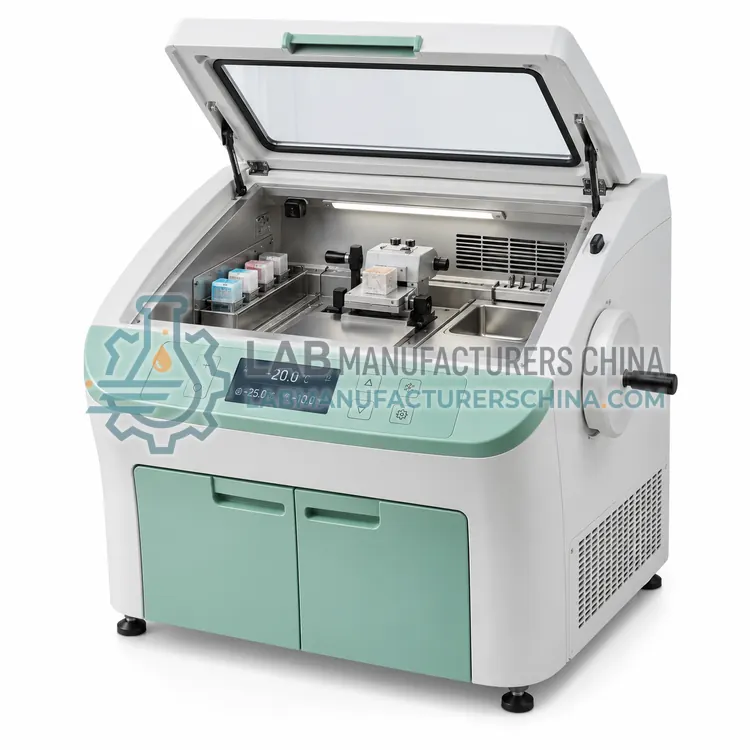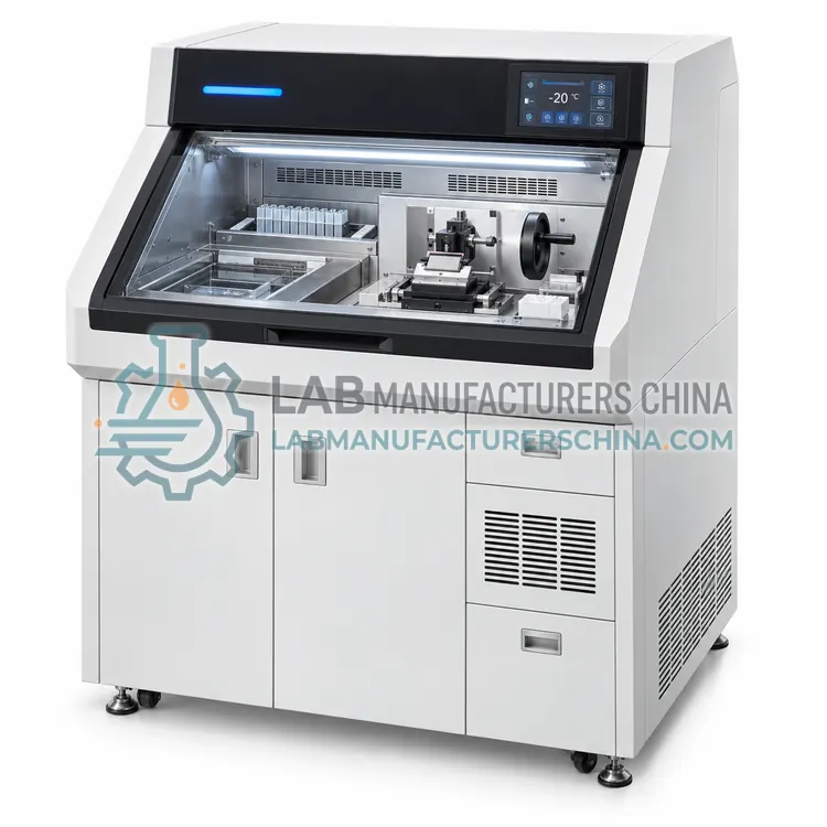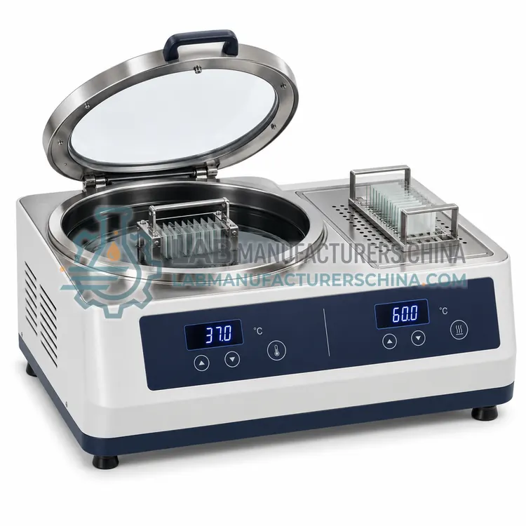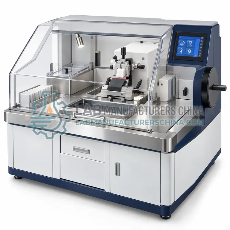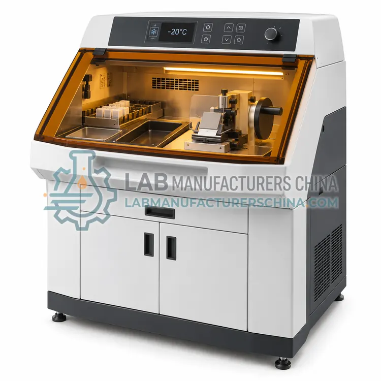
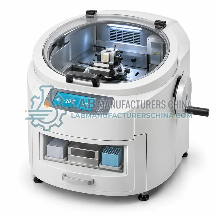
Research Cryostat
科研恒冷箱切片机
- SKU CRY-0002
- Cryostats
- CE (to be confirmed)
- IEC 61010-1 compliance (to be confirmed)
- IEC 61326-1 EMC (to be confirmed)
Price on request — FOB / CIF quoted per order
Prices depend on configuration, quantity and destination port.
Key specifications
| Chamber temperature range | –5 to –35 °C |
|---|---|
| Specimen temperature range | –10 to –40 °C |
| Section thickness range | 0.5–100 µm |
| Thickness increment | 0.5 µm below 5 µm |
| Cutting speed | 0–400 mm/s, adjustable |
| Cool-down time | about 2–3 h |
Request a quote Download brochure · to be provided
By Lab Manufacturers China Technical TeamLast reviewed Standards: IEC 61010-1, IEC 61010-2-101, IEC 61326-1
What is the Research Cryostat?
A research cryostat offers a wide temperature range for both chamber and specimen, programmable defrost and flexible cutting parameters. It suits laboratories developing new protocols, immunofluorescence and molecular work on fresh-frozen tissue.
What is the Research Cryostat?
A research cryostat offers a wide temperature range for both chamber and specimen, programmable defrost and flexible cutting parameters. It suits laboratories developing new protocols, immunofluorescence and molecular work on fresh-frozen tissue.
Overview
The research cryostat emphasises control and range rather than only speed. Chamber temperature can usually be set from about –5 to –35 °C, and the specimen stage is independently cooled over a similar or wider span, so the operator can make the block harder or softer than its surroundings. Section thickness may go below 1 µm for thin cryosections and up to 100 µm for free-floating work. Programmable defrost, adjustable cutting speed and fine anti-roll plate control support delicate protocols. Accessories may include a cryo-transfer system, a stereotaxic or pin-type holder and a light in the chamber. Researchers use these instruments for immunofluorescence, laser capture microdissection preparation, RNA work and neuroscience, where preservation of antigen and nucleic acid integrity matters.
How does it work?
A cryostat places a microtome inside an insulated chamber cooled by one or two refrigeration compressors. The chamber and often a separate specimen stage are brought to between –15 and –35 °C, freezing the tissue to a suitable hardness. The operator attaches the frozen block to a chuck with embedding medium and sections it with a disposable blade held at a fixed angle, producing sections typically 1 to 100 µm thick. Sections adhere to room-temperature slides and are fixed immediately in alcohol or acetone. Anti-roll plates and brush techniques guide ribbons free of curling. Because the whole cutting zone stays cold, tissue morphology is preserved without paraffin infiltration, and total turnaround from specimen to stained slide can be under 20 minutes.
What are the key features?
- Chamber temperature range about –5 to –35 °C
- Independently controlled specimen cooling
- Section thickness from below 1 µm to 100 µm
- Programmable defrost and adjustable cutting speed
- Fine anti-roll plate and chamber illumination
- Accessories for cryo-transfer and specialised holders
What are the technical specifications?
A typical research cryostat configuration offers chamber temperature range of –5 to –35 °C, specimen temperature range of –10 to –40 °C, section thickness range of 0.5–100 µm, thickness increment of 0.5 µm below 5 µm, cutting speed of 0–400 mm/s, adjustable and cool-down time of about 2–3 h. Values are indicative of the product class; the exact figures for each configuration are confirmed on the quotation and the calibration documents supplied with the instrument.
Which applications is it used for?
The Research Cryostat is used in immunofluorescence on fresh-frozen tissue, laser capture microdissection preparation, RNA and nucleic acid work, neuroscience and stereology and method development. Across the wider Cryostats class, laboratories also deploy it for intraoperative frozen sections, immunofluorescence on fresh tissue and enzyme histochemistry.
Which standards and certifications apply?
Relevant standards and methods include IEC 61010-1, IEC 61010-2-101, IEC 61326-1, ISO 15189 and ISO 13485. Certification candidates for this model are CE, IEC 61010-1 compliance and IEC 61326-1 EMC; each is to be confirmed for the specific configuration before shipment, and certificates are supplied only where they have been issued.
Place the cryostat in a room kept below 25 °C with good ventilation, because condensers reject significant heat. Allow several hours for initial cool-down, then verify chamber and specimen temperatures against a calibrated thermometer. Defrost and clean the chamber on schedule; residual cryo-embedding medium degrades section quality. Use anti-roll plates and change blades often. Monitor for frost build-up, compressor noise and door seal condition, and service annually. Maintain temperature logs, cleaning records and staff training, since these documents support accreditation of frozen section services.
How do I select the right Research Cryostat?
Specify a research cryostat when protocols require wide, independent temperature control, very thin or very thick sections, or special holders for cryo-transfer. It is more expensive than a clinical model but far more adaptable. State chamber and specimen temperature ranges, section thickness limits, speed range, holder types and required accessories.
How does it compare with other Cryostats variants?
Choose a cryostat by the work it must support. Clinical laboratories doing intraoperative frozen sections need fast cool-down, a stable chamber near –20 °C, reliable blade cooling and often UV sterilisation for contamination control. Research and immunohistochemistry laboratories value accurate independent specimen cooling, wide temperature range to –35 °С and a motorised or ergonomic cutting system for repetitive work. Confirm chamber and specimen temperature ranges, cool-down time, defrost cycle, section thickness range, blade type and whether the cutting area can be cleaned with the chamber at working temperature. Also check section transfer options and the footprint of the lab bench.
Full specifications
| Chamber temperature range | –5 to –35 °C |
|---|---|
| Specimen temperature range | –10 to –40 °C |
| Section thickness range | 0.5–100 µm |
| Thickness increment | 0.5 µm below 5 µm |
| Cutting speed | 0–400 mm/s, adjustable |
| Cool-down time | about 2–3 h |
| Blade type | Disposable low-profile or high-profile |
| Defrost | Programmable, timed |
| Power supply | 220 V, 50 Hz, about 1.2 kW |
Indicative values for this product class. Final specifications are confirmed on the quotation and in the calibration documents supplied with the instrument.
Applications
- Immunofluorescence on fresh-frozen tissue
- Laser capture microdissection preparation
- RNA and nucleic acid work
- Neuroscience and stereology
- Method development
Key features
- Chamber temperature range about –5 to –35 °C
- Independently controlled specimen cooling
- Section thickness from below 1 µm to 100 µm
- Programmable defrost and adjustable cutting speed
- Fine anti-roll plate and chamber illumination
- Accessories for cryo-transfer and specialised holders
Frequently asked questions about Research Cryostat
Because optimal cutting temperature depends on tissue type and is often different from the chamber setpoint. Independent control lets the operator freeze a dense block harder or keep delicate tissue warmer without changing the whole chamber condition.
Research cryostats can cut sections below 1 µm with careful setup, although routine frozen sections are 5 to 20 µm. Achieving the thinnest sections requires a sharp blade, correct temperature for the tissue and slow, steady cutting.
Yes, it is capable of producing diagnostic frozen sections, but the extra adjustability and cost are not needed for routine intraoperative work. Many laboratories keep a research cryostat for special studies and a simpler clinical cryostat for urgent cases.
A cryostat cuts frozen tissue sections inside a refrigerated chamber. It is used for rapid intraoperative diagnosis, for immunohistochemistry and enzyme work that need unfixed tissue, and for research when native morphology or antigen preservation is important.
Most frozen sections are cut with the chamber between –18 and –25 °C, and the specimen stage matched or set a few degrees colder for dense tissue. Fatty or soft tissue may need –30 °C, while delicate tissue such as lymph node is often cut nearer –15 °C.
Shredding usually means the tissue or chamber is too cold, the block is too thick or the blade is blunt. Crumpling can indicate the tissue is too warm. Adjust chamber and specimen temperature, use a fresh blade, trim to size and cut at a slow, steady speed.
Related models