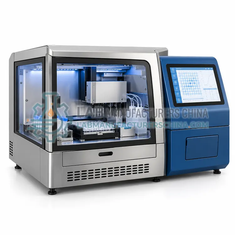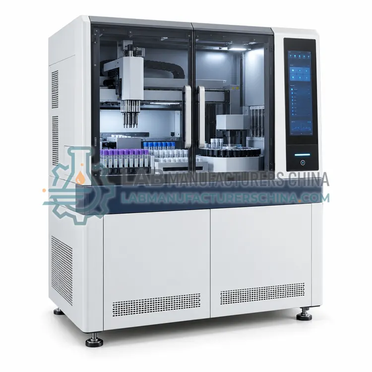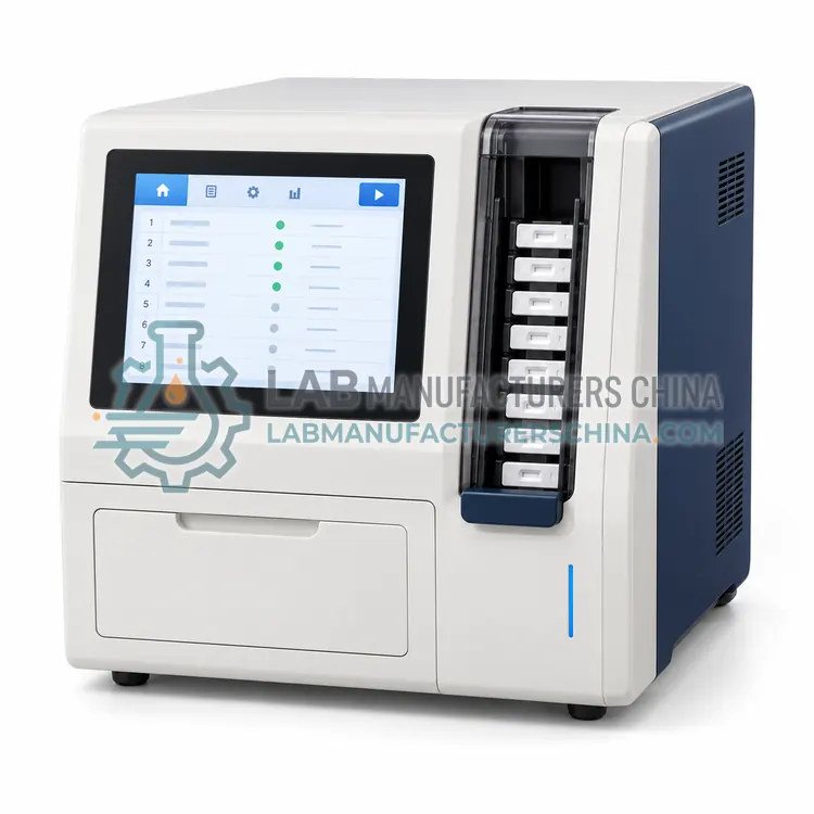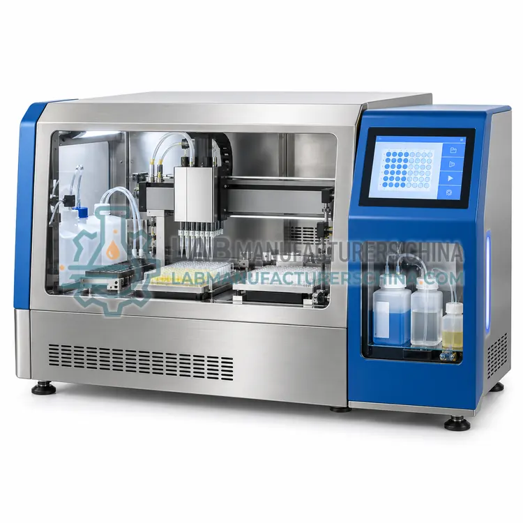
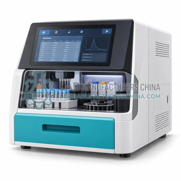
Magnetic Particle Chemiluminescence Immunoassay Analyzer
磁微粒化学发光免疫分析仪
- SKU CLI-0002
- CLIA Analyzers
- CE (to be confirmed)
- ISO 13485 manufacturing (to be confirmed)
- NMPA class II registration (to be confirmed)
Price on request — FOB / CIF quoted per order
Prices depend on configuration, quantity and destination port.
Key specifications
| Throughput | 150–300 tests/h |
|---|---|
| Solid phase | Paramagnetic microparticles, 1–3 µm |
| Detection | Enzymatic or flash chemiluminescence |
| Measuring range | Up to 4–5 orders of magnitude for selected assays |
| Separation cycles | 3–5 magnetic wash cycles |
| Sample volume | 20–100 µL per test |
Request a quote Download brochure · to be provided
By Lab Manufacturers China Technical TeamLast reviewed Standards: ISO 15189, CLSI EP05, CLSI EP15
What is the Magnetic Particle Chemiluminescence Immunoassay Analyzer?
This analyzer uses paramagnetic microparticles as the solid phase so that immune complexes can be captured and washed in a magnetic field, giving fast separation and low background. It suits laboratories that need high sensitivity for low-concentration markers across a wide menu.
What is the Magnetic Particle Chemiluminescence Immunoassay Analyzer?
This analyzer uses paramagnetic microparticles as the solid phase so that immune complexes can be captured and washed in a magnetic field, giving fast separation and low background. It suits laboratories that need high sensitivity for low-concentration markers across a wide menu.
Overview
Paramagnetic microparticles coated with capture antibody are mixed with sample and labelled detection reagent in a reaction cuvette. After incubation the magnet attracts the particles to the wall, unbound material is aspirated away and the particles are resuspended, and this cycle repeats before trigger solutions are added. Because separation is driven by a magnet rather than by centrifugation or filtration, the process is fast and requires little mechanical movement. Particle uniformity and lot control are important for precision, and the analyzer monitors agitation to keep particles suspended in the reagent bottle. The format gives a wide measuring range, suits sandwich assays for large molecules and competitive assays for small molecules, and typically delivers 150 to 300 tests per hour.
How does it work?
In chemiluminescence immunoassay the label is a molecule, often an acridinium ester or a luminol derivative, that emits light when triggered by an oxidant or a catalyst. Capture antibody or antigen is immobilised on magnetic particles, a well or a tube. After sample binding and washing, the labelled detection reagent forms a sandwich or competes with the analyte, and a trigger solution initiates the light-emitting reaction. A photomultiplier tube measures the light flash, which is integrated over a fixed interval, and the signal is converted into concentration using a calibration curve, usually a four-parameter logistic or a master curve with a lot-specific adjustment. Because light emission is measured against a dark background rather than as a difference between two absorbance values, chemiluminescence gives a wider dynamic range and better sensitivity than chromogenic enzyme immunoassay, and magnetic separation simplifies washing and automation.
What are the key features?
- Paramagnetic microparticles coated with capture antibody
- Magnetic capture and washing without centrifugation
- Continuous reagent agitation to keep particles suspended
- Wide measuring range for sandwich and competitive assays
- Low background for low-concentration analytes
- Particle lot control with onboard traceability
What are the technical specifications?
A typical magnetic particle chemiluminescence immunoassay analyzer configuration offers throughput of 150–300 tests/h, solid phase of Paramagnetic microparticles, 1–3 µm, detection of Enzymatic or flash chemiluminescence, measuring range of Up to 4–5 orders of magnitude for selected assays, separation cycles of 3–5 magnetic wash cycles and sample volume of 20–100 µL per test. Values are indicative of the product class; the exact figures for each configuration are confirmed on the quotation and the calibration documents supplied with the instrument.
Which applications is it used for?
The Magnetic Particle Chemiluminescence Immunoassay Analyzer is used in high-sensitivity hormone assays, cardiac and sepsis markers, infectious disease antigen detection and allergy and autoimmune testing. Across the wider CLIA Analyzers class, laboratories also deploy it for hormone and fertility testing, cardiac marker measurement in emergency care and tumour marker monitoring.
Which standards and certifications apply?
Relevant standards and methods include ISO 15189, CLSI EP05, CLSI EP15, CLSI EP17, CLSI I/LA20, IEC 61010-1 and IEC 61010-2-101. Certification candidates for this model are CE, ISO 13485 manufacturing and NMPA class II registration; each is to be confirmed for the specific configuration before shipment, and certificates are supplied only where they have been issued.
Install the analyzer on a stable bench with controlled temperature and humidity, a grounded power supply and space for reagent and waste containers. Verify performance at installation with precision, linearity and method comparison studies for every assay you will report, following CLSI EP05, EP15 and EP17 where detection limits are relevant. Establish calibration and control intervals per assay and document the curve validity period. Daily work includes checking reagent volumes and waste, running controls and reviewing the calibration status; trigger solutions and wash buffers must be prepared or replaced as specified because deteriorated solutions cause erratic light signals. Replace tubing, syringes and photodetector-related parts on schedule, and treat all samples, controls and liquid waste as biohazardous.
How do I select the right Magnetic Particle Chemiluminescence Immunoassay Analyzer?
Specify this variant when low detection limits and a wide dynamic range matter for your menu, and when throughput above about one hundred and fifty tests per hour is required. In the RFQ ask for functional sensitivity at the clinical decision point of each key analyte, particle lot consistency data, and the calibration stability for the assays you will report most often.
How does it compare with other CLIA Analyzers variants?
Select a CLIA analyzer from the assay menu first, because registration of each test with the platform is the decisive constraint, then size throughput from daily and peak-hour demand plus about thirty percent headroom. Check the first-result time for emergency markers such as troponin and beta-hCG, since several platforms need twenty to thirty minutes. Compare sensitivity and linear range for the analytes that matter to your clinical service, the sample volume per test, and the calibration stability, which determines how often curves must be rebuilt. Decide between magnetic bead, coated tube and microplate formats according to menu size and acceptable hands-on time. Confirm onboard reagent capacity, refrigeration, STAT access, carryover control, LIS interface, and the availability of calibrators, controls and service support locally.
Full specifications
| Throughput | 150–300 tests/h |
|---|---|
| Solid phase | Paramagnetic microparticles, 1–3 µm |
| Detection | Enzymatic or flash chemiluminescence |
| Measuring range | Up to 4–5 orders of magnitude for selected assays |
| Separation cycles | 3–5 magnetic wash cycles |
| Sample volume | 20–100 µL per test |
| Reagent agitation | Continuous orbital mixing onboard |
| Calibration stability | 21–28 days depending on the assay |
| Interfaces | LAN with HL7 or ASTM, USB |
Indicative values for this product class. Final specifications are confirmed on the quotation and in the calibration documents supplied with the instrument.
Applications
- High-sensitivity hormone assays
- Cardiac and sepsis markers
- Infectious disease antigen detection
- Allergy and autoimmune testing
Key features
- Paramagnetic microparticles coated with capture antibody
- Magnetic capture and washing without centrifugation
- Continuous reagent agitation to keep particles suspended
- Wide measuring range for sandwich and competitive assays
- Low background for low-concentration analytes
- Particle lot control with onboard traceability
Frequently asked questions about Magnetic Particle Chemiluminescence Immunoassay Analyzer
Particles provide a much larger surface area in suspension, so binding is faster and washing is more efficient, which lowers background and improves sensitivity. Magnetic capture also allows the solid phase to be separated without centrifugation, which is quicker and easier to automate.
If particles settle in the reagent bottle, the concentration dispensed varies and results drift. Analyzers therefore agitate reagents continuously or before aspiration, and the laboratory should check that mixing is operating whenever precision problems appear.
Both sandwich assays for large analytes such as proteins and hormones and competitive assays for small molecules such as therapeutic drugs and some hormones. Competitive assays need different curve models and control material, so confirm that the platform and software support the formats in your menu.
Functional sensitivity is the lowest concentration measurable with acceptable precision, often expressed as the concentration with a coefficient of variation of twenty percent. It should be verified locally with repeated measurements of low-level samples, because the manufacturer value may differ from performance in your laboratory.
Chemiluminescence measures light emission against a dark background, giving a wider measuring range and lower detection limits than the absorbance difference measured in ELISA. It also automates more easily because magnetic separation replaces repeated plate washing, though reagent and instrument costs are usually higher.
Most assays use a stored master curve with a lot-specific calibration that remains valid for two to four weeks, with a full curve required at each new reagent lot or after major maintenance. Daily control results confirm that the calibration remains acceptable, and any shift triggers recalibration before reporting.
At extremely high analyte concentrations both capture and detection antibodies become saturated and the signal falls, which can produce a falsely low result. Most analyzers detect this by diluting and re-measuring samples above a defined concentration or by comparing the reaction rate with expected curve behaviour.
Related models