Cytocentrifuge
细胞离心机
- SKU CLC-0004
- Clinical Centrifuges
- CE (to be confirmed)
- ISO 13485 quality system (to be confirmed)
Price on request — FOB / CIF quoted per order
Prices depend on configuration, quantity and destination port.
Key specifications
| Speed range | 100–2,000 rpm |
|---|---|
| Maximum RCF | Approx. 800–1,000 × g |
| Capacity | 6–12 slides per run |
| Sample volume | 100–500 µL per chamber |
| Deposition area | Approx. 6–25 mm diameter |
| Timer | 1–99 min |
Request a quote Download brochure · to be provided
By Lab Manufacturers China Technical TeamLast reviewed Standards: IEC 61010-2-020, IEC 61010-1, ISO 15189
What is the Cytocentrifuge?
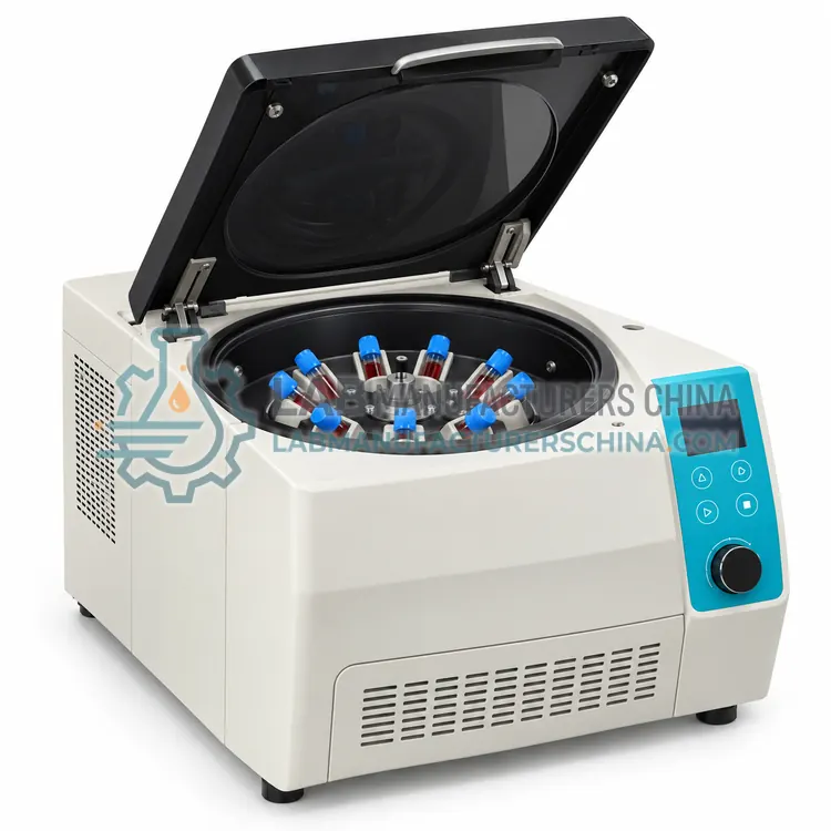
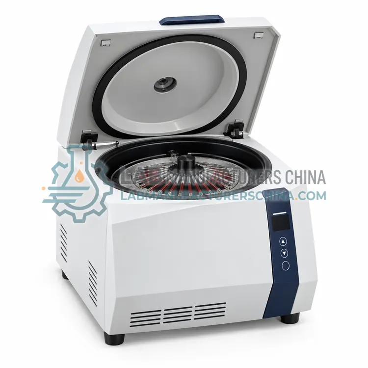
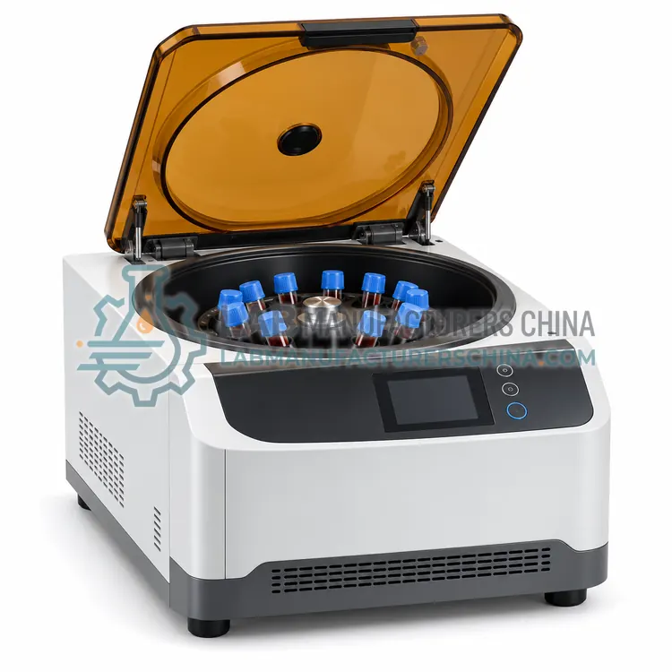
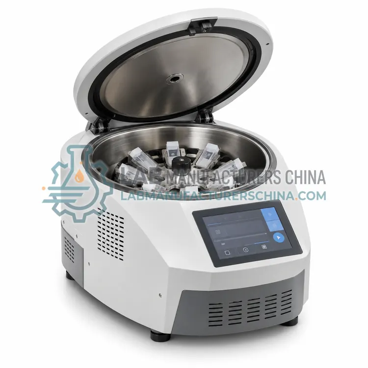
A cytocentrifuge deposits cells from low-cell-count fluids such as cerebrospinal fluid, body fluids and bronchoalveolar lavage onto a glass slide within a small circular area using gentle force. It produces flattened monolayers for microscopic differential counting and immunocytochemistry.
What is the Cytocentrifuge?
A cytocentrifuge deposits cells from low-cell-count fluids such as cerebrospinal fluid, body fluids and bronchoalveolar lavage onto a glass slide within a small circular area using gentle force. It produces flattened monolayers for microscopic differential counting and immunocytochemistry.
Overview
In a cytocentrifuge, a sample chamber is clamped to a glass slide with a filter card between them. Spinning at low speed, typically 500 to 2,000 rpm for 3 to 10 minutes, forces cells through the chamber opening onto a circular spot of 6 to 25 mm, while the filter card absorbs the fluid. The result is a thin monolayer with good morphology, where as few as 100 to 500 cells per sample can be concentrated. Rotors hold 6 to 12 chambers and some models offer programmable acceleration to limit cell damage. Slides are air-dried and stained with Wright-Giemsa, Papanicolaou or immunocytochemical methods. Disposable funnels, filter cards and clips are consumables, and washable aerosol-tight lids protect staff from aerosols.
How does it work?
A centrifuge replaces gravity with a much larger centrifugal force. A motor, usually a brushless induction or DC drive, spins a rotor holding sample tubes, and relative centrifugal force rises with the square of speed: RCF = 1.118 × 10⁻⁵ × r × rpm², where r is radius in centimeters. Heavier components such as red cells move to the tube bottom while serum or plasma stays above, often separated by a gel barrier that forms a solid layer during spinning. Swing-out rotors let tubes settle horizontally for a flat interface, while fixed-angle rotors shorten pelleting distance. Refrigerated models use a compressor to hold chamber temperature at 4 to 20 °C, and safety systems include lid locks, imbalance detection and overspeed protection. Programmable acceleration and braking profiles prevent remixing of separated layers.
What are the key features?
- Deposits cells in a 6–25 mm circular area on glass slides
- Low speed 500–2,000 rpm to protect cell morphology
- Rotor for 6 to 12 sample chambers per run
- Programmable acceleration and time settings
- Filter cards absorb fluid while cells adhere to the slide
- Aerosol-tight lid for biosafety
What are the technical specifications?
A typical cytocentrifuge configuration offers speed range of 100–2,000 rpm, maximum RCF of Approx. 800–1,000 × g, capacity of 6–12 slides per run, sample volume of 100–500 µL per chamber, deposition area of Approx. 6–25 mm diameter and timer of 1–99 min. Values are indicative of the product class; the exact figures for each configuration are confirmed on the quotation and the calibration documents supplied with the instrument.
Which applications is it used for?
The Cytocentrifuge is used in cerebrospinal fluid cell differential, body fluid cytology, bronchoalveolar lavage analysis and immunocytochemistry slide preparation. Across the wider Clinical Centrifuges class, laboratories also deploy it for serum and plasma separation, blood component preparation and microhematocrit determination.
Which standards and certifications apply?
Relevant standards and methods include IEC 61010-2-020, IEC 61010-1, ISO 15189, CLSI GP44, ISO 13485 and EN 12469. Certification candidates for this model are CE and ISO 13485 quality system; each is to be confirmed for the specific configuration before shipment, and certificates are supplied only where they have been issued.
Place the centrifuge on a rigid level bench with clearance around it, and never run it unbalanced; opposite tubes must match within the tolerance stated in the manual. Use sealed buckets or aerosol-tight lids for infectious specimens. Verify speed with a photo-tachometer and temperature with a reference thermometer during installation and annually. Inspect rotors for cracks, corrosion and pitting, record run hours, and retire them at the manufacturer's service life. Clean with mild disinfectant, and clean the condenser of refrigerated models. Keep records under ISO 15189 and follow IEC 61010-2-020 safety requirements.
How do I select the right Cytocentrifuge?
Select this variant when low-cellularity samples must be concentrated into a stained monolayer, especially CSF and serous fluids. In the RFQ specify the number of chambers, speed and acceleration control, deposition area, slide and filter card format, and supply of disposable chambers. Ask which stain workflows the supplier has validated.
How does it compare with other Clinical Centrifuges variants?
Choose by sample type, tube format and temperature need. Routine serum or plasma separation in gel tubes needs a swing-out rotor reaching 3,000–4,000 rpm, roughly 1,500 to 2,200 × g, and a capacity matching peak hourly tube load. Blood bank and platelet work call for refrigeration at 20–24 °C or 4 °C and larger bucket capacity. Microhematocrit and cytospin tasks need purpose-built rotors. Check rotor and adapter compatibility with your tube diameters, maximum RCF, noise, footprint and programmability of acceleration and braking. Confirm lid lock and imbalance safety, biohazard containment with sealed buckets, and availability of spare rotors and service.
Full specifications
| Speed range | 100–2,000 rpm |
|---|---|
| Maximum RCF | Approx. 800–1,000 × g |
| Capacity | 6–12 slides per run |
| Sample volume | 100–500 µL per chamber |
| Deposition area | Approx. 6–25 mm diameter |
| Timer | 1–99 min |
| Acceleration | Programmable, 1–9 steps |
| Slide format | Standard 25 × 75 mm microscope slides |
| Lid | Aerosol-tight, interlocked |
| Noise level | ≤ 60 dB(A) |
| Power supply | 220 V AC, 50/60 Hz, 150–250 W |
Indicative values for this product class. Final specifications are confirmed on the quotation and in the calibration documents supplied with the instrument.
Applications
- Cerebrospinal fluid cell differential
- Body fluid cytology
- Bronchoalveolar lavage analysis
- Immunocytochemistry slide preparation
Key features
- Deposits cells in a 6–25 mm circular area on glass slides
- Low speed 500–2,000 rpm to protect cell morphology
- Rotor for 6 to 12 sample chambers per run
- Programmable acceleration and time settings
- Filter cards absorb fluid while cells adhere to the slide
- Aerosol-tight lid for biosafety
Frequently asked questions about Cytocentrifuge
It concentrates cells from fluids with few cells onto a glass slide using gentle centrifugal force. The cells form a thin, flat monolayer that can be stained and examined under a microscope, making it possible to classify white cells or identify abnormal cells in CSF and body fluids.
The filter card absorbs the liquid that passes through the chamber opening, so cells are retained and flattened on the slide rather than washed off. Card type and weight influence cell recovery and morphology, so use the card specified for your chamber.
Typical settings are 500 to 1,000 rpm for 5 to 10 minutes for CSF and fluids, with 1,500 to 2,000 rpm for some applications. Lower speed and gradual acceleration protect fragile cells, and each laboratory validates settings for its specimen types.
It separates blood and other specimens by density. Typical uses are obtaining serum or plasma before analysis, packing red cells for hematocrit, preparing blood components in blood banks, and concentrating cells onto slides for cytology. It is a core pre-analytical instrument in laboratories.
Rpm is rotor speed in revolutions per minute, while RCF is the actual force on the sample, expressed in multiples of gravity. RCF depends on both speed and rotor radius, so protocols should state RCF in × g to give the same separation on different centrifuges.
Refrigeration limits heating from rotor friction and preserves temperature-sensitive analytes or cells. Blood bank platelet preparations are spun near 20–24 °C, while plasma and some hormone or enzyme samples are spun at 4 °C. Unrefrigerated units can warm by several degrees during long runs.