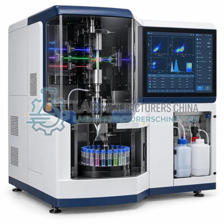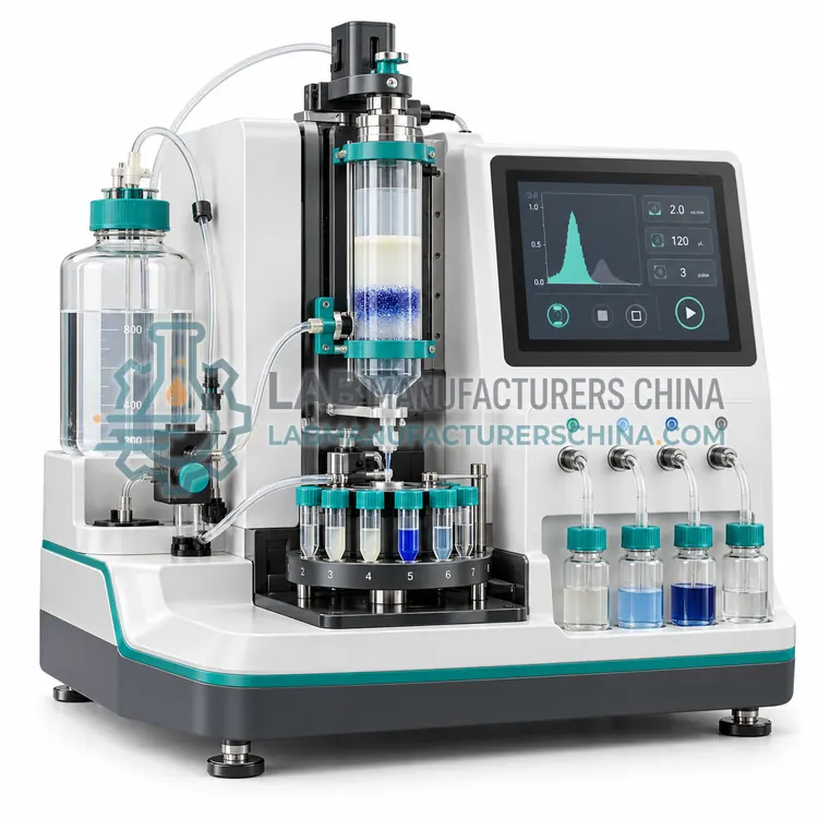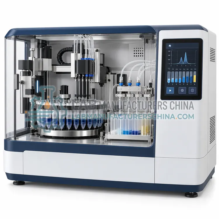
Microfluidic Cell Sorter
微流控细胞分选仪
- SKU CES-0003
- Cell Separation
- CE (to be confirmed)
- ISO 9001 manufacturing (to be confirmed)
- IEC 61010-1 safety (to be confirmed)
Price on request — FOB / CIF quoted per order
Prices depend on configuration, quantity and destination port.
Key specifications
| Throughput | hundreds to a few thousand cells per second |
|---|---|
| Sample volume | 10 µL–5 mL |
| Channel size | approx. 50–300 µm |
| Cell size range | approx. 2–30 µm |
| Separation modes | Acoustic, optical or pressure switching |
| Purity | 80–95% (mode dependent) |
Request a quote Download brochure · to be provided
By Lab Manufacturers China Technical TeamLast reviewed Standards: ISO 20387, USP <1046>, IEC 61010-1
What is the Microfluidic Cell Sorter?
A microfluidic cell sorter deflects cells in narrow channels using acoustic waves, optical forces or fluid switching, isolating target populations from small samples with low shear stress and no aerosol generation, which suits delicate primary cells and limited clinical material.
What is the Microfluidic Cell Sorter?
A microfluidic cell sorter deflects cells in narrow channels using acoustic waves, optical forces or fluid switching, isolating target populations from small samples with low shear stress and no aerosol generation, which suits delicate primary cells and limited clinical material.
Overview
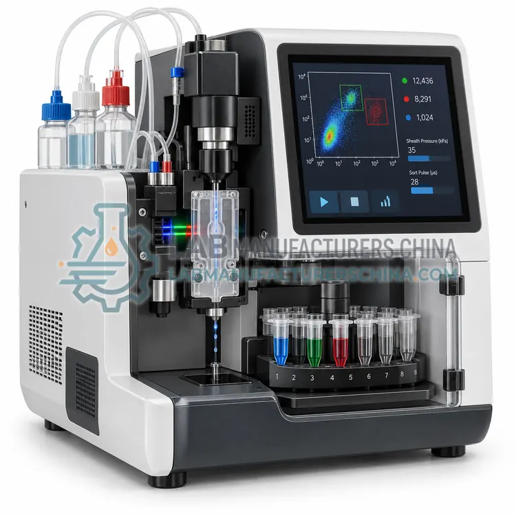
The device flows cells through a microchannel, often a single-use cartridge, where a transducer, laser or pressure pulse diverts selected cells into a collection channel. Acoustic systems use a standing wave that moves larger or denser cells toward a pressure node, which allows label-free separation by size and density. Optical and fluorescence-triggered systems label cells and deflect them when a signal is detected. Because channels are small, cells experience low shear stress and the dead volume is minimal, which suits low-input samples such as cerebrospinal fluid or fine-needle aspirates. Throughput is usually lower than droplet sorting, typically hundreds to a few thousand cells per second, and clogging risk makes filtration essential. Cartridges are disposable to prevent cross-contamination between samples.
How does it work?
Separation exploits a physical or biochemical difference between the target and the rest of the sample. Immunomagnetic methods coat cells with antibody-conjugated magnetic particles and either retain labelled cells in a column placed in a magnetic field, or label everything except the target and let the desired cells flow through untouched. Fluorescence-activated sorting stains cells with fluorescent antibodies, interrogates each cell in a fluid stream and applies a charge to the droplet containing a selected cell, which is then deflected into a collection tube. Microfluidic sorters use narrow channels, acoustic waves or optical forces to deflect cells without droplets. Density-based separation layers the sample over a medium of defined density so that cells of different density band at different interfaces during centrifugation. Each method trades purity, yield, speed and gentleness differently, and downstream assays determine which balance is acceptable.
What are the key features?
- Single-use microfluidic cartridge to prevent cross-contamination
- Acoustic, optical or pressure-based cell deflection
- Low shear stress suitable for sensitive primary cells
- Small dead volume for low-input samples
- Label-free size and density separation in acoustic mode
- Compact benchtop format with simple fluidic connections
What are the technical specifications?
A typical microfluidic cell sorter configuration offers throughput of hundreds to a few thousand cells per second, sample volume of 10 µL–5 mL, channel size of approx. 50–300 µm, cell size range of approx. 2–30 µm, separation modes of Acoustic, optical or pressure switching and purity of 80–95% (mode dependent). Values are indicative of the product class; the exact figures for each configuration are confirmed on the quotation and the calibration documents supplied with the instrument.
Which applications is it used for?
The Microfluidic Cell Sorter is used in sorting from low-volume clinical samples, label-free separation of blood cell types, enrichment before single-cell sequencing and separation of shear-sensitive primary cells. Across the wider Cell Separation class, laboratories also deploy it for T cell and immune subset isolation, stem cell and progenitor enrichment and cell therapy starting material preparation.
Which standards and certifications apply?
Relevant standards and methods include ISO 20387, USP <1046>, IEC 61010-1, IEC 61010-2-020, IEC 60825-1 and ISO 15189. Certification candidates for this model are CE, ISO 9001 manufacturing and IEC 61010-1 safety; each is to be confirmed for the specific configuration before shipment, and certificates are supplied only where they have been issued.
Work in a biosafety cabinet with appropriate containment, and keep separation buffers cold unless the protocol states otherwise, since cells are more sensitive to handling than to the method itself. Use fresh antibodies and beads, avoid vigorous pipetting, and filter samples through a 30–40 µm mesh to prevent column or nozzle blockage. Include a dead-cell removal step, because dead cells bind non-specifically and reduce purity. After separation, verify purity and viability by flow cytometry and record recovery and purity values for each preparation. Aseptic technique and closed systems are required for cell therapy workflows, where separation is part of a validated process under ISO 20387 and USP <1046> expectations, including documented operator training and traceability of reagents and lots.
How do I select the right Microfluidic Cell Sorter?
Select a microfluidic sorter when sample volume is limited, when shear sensitivity or aerosol containment is a concern, or when label-free separation is sufficient. Specify separation mode, throughput, cell size range, purity and the cartridge format and cost per sample.
How does it compare with other Cell Separation variants?
Choose by the required purity, yield, cell number and downstream application. Magnetic separation is fast, gentle and scalable, works with 10⁷ to 10⁹ cells and needs no sorter, but purity is typically 90–98%, so it may not be sufficient for single-cell sequencing. Fluorescence-activated sorting reaches 98–99.9% purity, can deposit single cells into plates with index information and supports multi-parameter gating, at the cost of lower throughput, higher shear stress and specialised operation. Microfluidic and acoustic methods suit low-volume samples and cell types sensitive to shear. Density gradient separation remains the standard first step for blood and bone marrow, followed by magnetic or flow-based purification. Confirm compatibility of labels, buffers and consumables with downstream culture, functional assays and regulatory requirements.
Full specifications
| Throughput | hundreds to a few thousand cells per second |
|---|---|
| Sample volume | 10 µL–5 mL |
| Channel size | approx. 50–300 µm |
| Cell size range | approx. 2–30 µm |
| Separation modes | Acoustic, optical or pressure switching |
| Purity | 80–95% (mode dependent) |
| Viability | above 90% (typical) |
| Cartridge | Single-use, sterilised |
| Aerosol generation | None, closed fluidic path |
| Dimensions (W x D x H) | approx. 300 x 400 x 300 mm |
Indicative values for this product class. Final specifications are confirmed on the quotation and in the calibration documents supplied with the instrument.
Applications
- Sorting from low-volume clinical samples
- Label-free separation of blood cell types
- Enrichment before single-cell sequencing
- Separation of shear-sensitive primary cells
Key features
- Single-use microfluidic cartridge to prevent cross-contamination
- Acoustic, optical or pressure-based cell deflection
- Low shear stress suitable for sensitive primary cells
- Small dead volume for low-input samples
- Label-free size and density separation in acoustic mode
- Compact benchtop format with simple fluidic connections
Frequently asked questions about Microfluidic Cell Sorter
Microfluidic sorters process hundreds to a few thousand cells per second, well below the 5,000–30,000 of droplet sorters. They compensate with lower shear, smaller sample requirements, no aerosol generation and simpler operation.
No. A standing acoustic wave moves cells according to size, density and compressibility, so populations can be separated without antibodies. Purity is lower than antibody-based methods but cells remain untouched for downstream culture.
Cell clumps, fibrin strands and debris larger than the channel are the main causes. Filtering through a 30–40 µm mesh and avoiding over-concentrated suspensions keeps the cartridge flowing reliably.
Positive selection labels the target cells so they are retained or collected. Negative selection labels everything except the target, so the desired cells remain untouched and unlabelled, which is preferred when the downstream assay is sensitive to bound antibody.
Typically 90–98% for positive selection and similar for well-optimised negative selection, depending on the marker and sample. Flow sorting raises purity to 98–99.9%, but yields fewer cells and takes longer.
Clumps and fibrin block magnetic columns and clog sorter nozzles, causing loss of sample and poor reproducibility. Passing the suspension through a 30–40 µm mesh removes aggregates while keeping single cells intact.
Related models