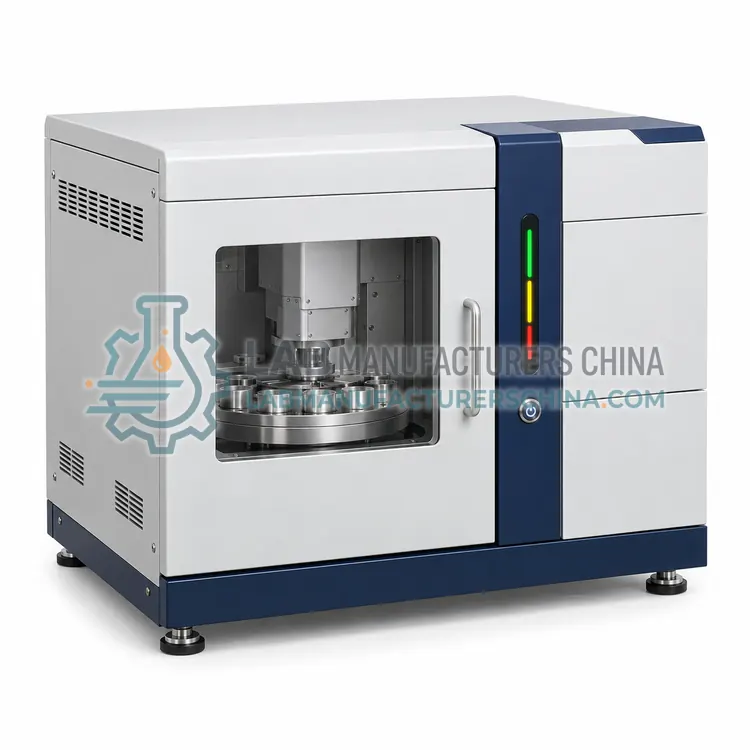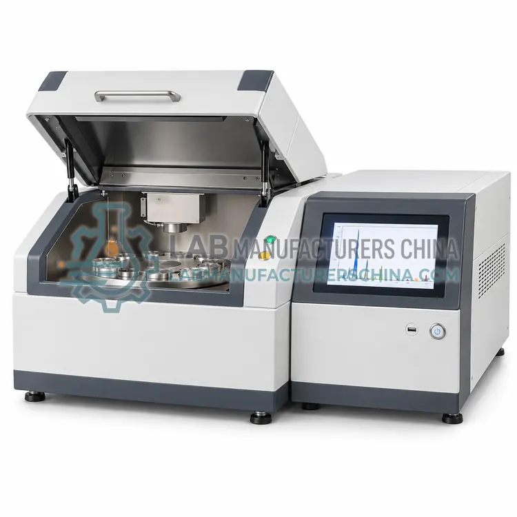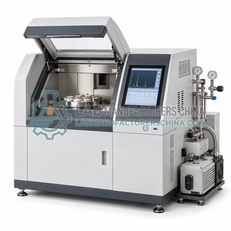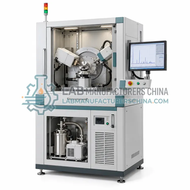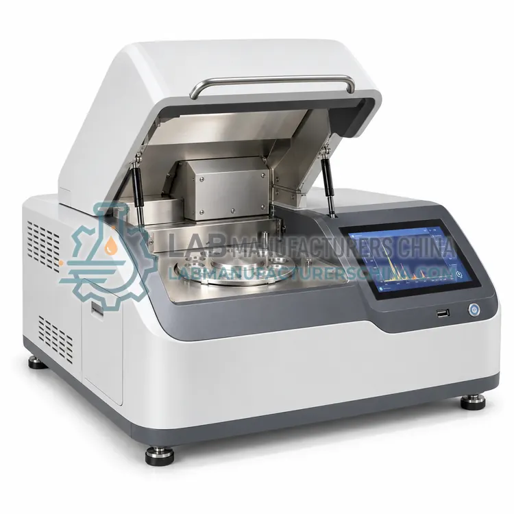
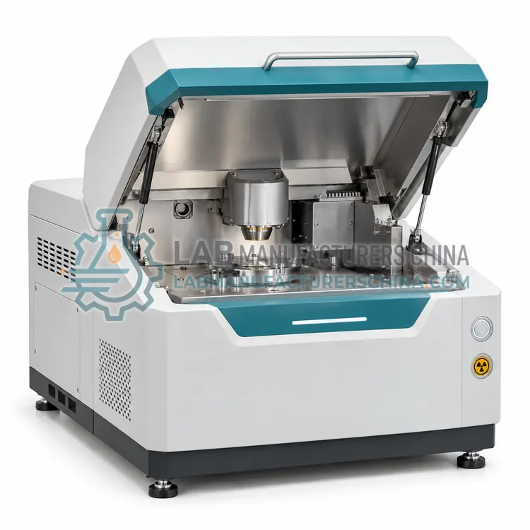
Benchtop Micro-XRF Spectrometer
台式微区 XRF 光谱仪
- SKU BEX-0004
- Benchtop XRF
- CE (to be confirmed)
- Radiation safety enclosure (to be confirmed)
- ISO 9001 manufacturing (to be confirmed)
Price on request — FOB / CIF quoted per order
Prices depend on configuration, quantity and destination port.
Key specifications
| Spot size | 10–100 µm |
|---|---|
| Element range | Na to U |
| Detection limit | 10–1000 mg/kg typical |
| Stage travel | up to 100 × 100 mm |
| Stage resolution | 1 µm |
| X-ray tube power | 30–100 W |
Request a quote Download brochure · to be provided
By Lab Manufacturers China Technical TeamLast reviewed Standards: ASTM E1621 Wavelength dispersive X-ray fluorescence, IEC 62321 Determination of restricted substances in electrotechnical products, ISO 3497 Metallic coatings — X-ray spectrometric thickness
What is the Benchtop Micro-XRF Spectrometer?
A benchtop micro-XRF spectrometer focuses the beam to a small spot for spatially resolved elemental mapping. It is chosen for small features, inclusions, coatings and contamination analysis in a compact instrument without a synchrotron.
What is the Benchtop Micro-XRF Spectrometer?
A benchtop micro-XRF spectrometer focuses the beam to a small spot for spatially resolved elemental mapping. It is chosen for small features, inclusions, coatings and contamination analysis in a compact instrument without a synchrotron.
Overview
Polycapillary optics concentrate X-rays from the tube onto a spot of roughly 10 to 100 micrometres, and a motorized stage moves the sample to build maps and line scans. This brings micro-analysis capability into a benchtop footprint without a synchrotron or electron microscope. It is used to examine inclusions in minerals, particles and contamination, solder joints and electronics, coating layers and cultural heritage objects, all without sample damage. Sensitivity per element is lower than large-spot XRF because the beam is concentrated, so measurement times are longer. Vacuum or helium purge improves light-element detection, and stage reproducibility determines map accuracy. The analysis is a natural complement to optical or electron microscopy when elemental identity is needed.
How does it work?
A modest-power X-ray tube excites the sample inside a shielded enclosure, and a detector measures the characteristic X-rays emitted by the elements present. Energy-dispersive benchtop models use a silicon drift detector and record the full spectrum simultaneously, which gives fast measurement and a compact design. Wavelength-dispersive benchtop models use analyzing crystals and give better resolution and lower detection limits at higher cost. Vacuum or helium purge extends the element range toward sodium, and filters or secondary targets reduce background. The result is processed with fundamental parameters or empirical calibrations matched to the sample matrix. Enclosed geometry and interlocks ensure that no measurable radiation escapes during normal operation, and sample changers allow unattended sequences.
What are the key features?
- Polycapillary optics with 10 to 100 µm spot
- Motorized stage for maps and line scans
- Non-destructive analysis of small features
- Inclusion, particle and coating analysis
- Vacuum or helium purge for light elements
- Complement to optical and electron microscopy
What are the technical specifications?
A typical benchtop micro-xrf spectrometer configuration offers spot size of 10–100 µm, element range of Na to U, detection limit of 10–1000 mg/kg typical, stage travel of up to 100 × 100 mm, stage resolution of 1 µm and X-ray tube power of 30–100 W. Values are indicative of the product class; the exact figures for each configuration are confirmed on the quotation and the calibration documents supplied with the instrument.
Which applications is it used for?
The Benchtop Micro-XRF Spectrometer is used in mineral inclusions and geological phases, coating, solder and electronics analysis, contamination particles and failure analysis and cultural heritage and archaeological objects. Across the wider Benchtop XRF class, laboratories also deploy it for alloy and incoming material verification, roHS and restricted substance screening and cement, glass and ceramic analysis.
Which standards and certifications apply?
Relevant standards and methods include ASTM E1621 Wavelength dispersive X-ray fluorescence, IEC 62321 Determination of restricted substances in electrotechnical products, ISO 3497 Metallic coatings — X-ray spectrometric thickness, ASTM D4294 Sulfur in petroleum products by EDXRF, IEC 61010-2-091 and ISO/IEC 17025. Certification candidates for this model are CE, Radiation safety enclosure and ISO 9001 manufacturing; each is to be confirmed for the specific configuration before shipment, and certificates are supplied only where they have been issued.
Enclosed benchtop instruments are radiation devices, so installation requires correct shielding, functional interlocks, radiation survey and operator training. Sample size and thickness must fit the enclosure and the analytical requirement, and powders are best pressed into pellets for reproducibility. Matrix-matched calibration is essential for accuracy, with drift monitored by check standards. Vacuum pumps, filters and X-ray tubes are consumable and wear items. Avoid measuring liquids in unsupported open cups, and use sample spinners or masks where the software recommends them. Keep service and calibration records, and dispose of tubes and detectors according to radiation regulations.
How do I select the right Benchtop Micro-XRF Spectrometer?
Select benchtop micro-XRF when elemental maps of small features are needed and the sample must remain intact, but a synchrotron or electron microscope is not available. Specify the spot size, map area and element range. Confirm stage reproducibility and whether vacuum or helium is required for light elements.
How does it compare with other Benchtop XRF variants?
Select a benchtop XRF configuration by the element range, required detection limits and sample types. Energy-dispersive models are the most common and cost-effective for screening, alloy and material verification and multi-element quantification. Wavelength-dispersive benchtop models suit light elements and more precise analysis when space and budget allow. Add vacuum or helium purge for light elements, and choose application calibrations for RoHS, cement, polymers or catalysts. Micro-XRF versions add spatial resolution for small features and coatings. Consider sample size limits inside the enclosure, the number of sample positions, data export and regulatory methods. Confirm whether a special calibration or reference material set is needed before ordering.
Full specifications
| Spot size | 10–100 µm |
|---|---|
| Element range | Na to U |
| Detection limit | 10–1000 mg/kg typical |
| Stage travel | up to 100 × 100 mm |
| Stage resolution | 1 µm |
| X-ray tube power | 30–100 W |
| Detector | silicon drift detector |
| Mapping | point, line and area maps |
| Enclosure | shielded with interlocks |
| Interface | Ethernet |
Indicative values for this product class. Final specifications are confirmed on the quotation and in the calibration documents supplied with the instrument.
Applications
- Mineral inclusions and geological phases
- Coating, solder and electronics analysis
- Contamination particles and failure analysis
- Cultural heritage and archaeological objects
Key features
- Polycapillary optics with 10 to 100 µm spot
- Motorized stage for maps and line scans
- Non-destructive analysis of small features
- Inclusion, particle and coating analysis
- Vacuum or helium purge for light elements
- Complement to optical and electron microscopy
Frequently asked questions about Benchtop Micro-XRF Spectrometer
Micro-XRF has lower spatial resolution but works in air, requires no conductive coating and penetrates deeper, which suits insulating, hydrated and layered samples. SEM-EDS resolves much finer features under vacuum. Many laboratories use both, choosing by required resolution, sample state and depth of interest.
Yes, if the inclusion is larger than the beam spot and can be located on the stage. Smaller inclusions are measured only partially, and the result is influenced by the surrounding matrix. Line scans and maps across the inclusion help interpret the data and confirm its boundaries.
Concentrating the beam onto a small spot reduces the total photon flux reaching the sample and, for maps, divides the measurement across many pixels. Each point therefore needs a longer dwell time to collect enough counts for reliable results, increasing total measurement time.
The sample and primary beam are fully enclosed, and interlocks prevent operation with the lid open, so no measurable radiation escapes during normal use. Some instruments emit a small amount when the shutter opens briefly or when disabled for service. Radiation surveys and dosimetry are used where local rules require them.
Benchtop enclosures typically accept samples from a few millimetres up to about 100 to 300 millimetres across, depending on the model. Larger parts must be cut or measured with a portable analyzer. Powder cups, pressed pellets and thin films are the most common sample forms in benchtop systems.
Yes, liquids are measured in cups with thin film windows, and volatile or reactive liquids require special cups and venting. Light elements in liquids are difficult because of absorption and evaporation. Some matrices are better measured after digestion or as dried residues on a carrier.
Related models