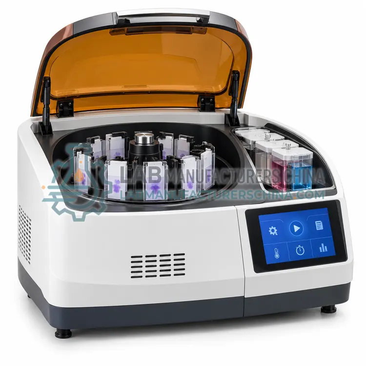
How do Cytocentrifuges work?
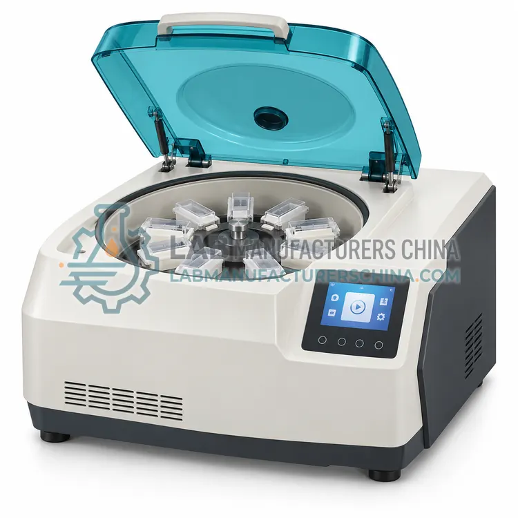
A cytocentrifuge is a low-speed centrifuge fitted with sample chambers that hold a microscope slide against a card or gasket. The fluid sample is placed in the chamber and centrifuged, so cells move out of suspension and settle onto the slide within a defined circle of typically 6 to 13 mm. Absorbent card or filter paper beside the slide wicks away the supernatant, leaving cells concentrated in a monolayer with minimal protein background. Spin speed and time, usually 500 to 1500 rpm for 3 to 10 minutes, control cell density and flattening. Faster spins pack more cells but flatten them and increase background; slower spins preserve nuclear detail. Some instruments add a cytology funnel, a slide clip and a disassembly system that protects the operator from aerosols.
Which Cytocentrifuges variants are available?
- Single-Chamber Cytocentrifuge — A single-chamber cytocentrifuge processes one sample at a time and is the most economical option.
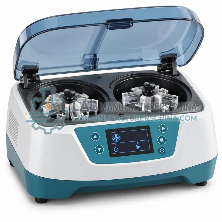
- Double-Chamber Cytocentrifuge — A double-chamber cytocentrifuge prepares two samples in one run, roughly doubling throughput while keeping the same bench footprint.
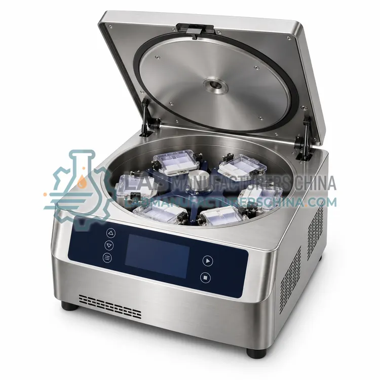
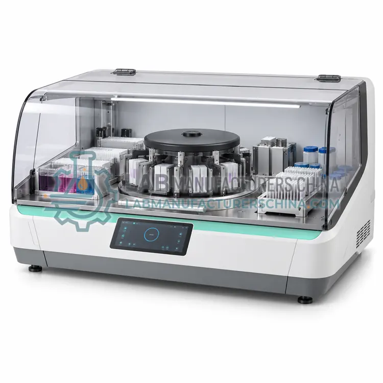
- Multi-Chamber Cytocentrifuge — A multi-chamber cytocentrifuge holds several sample chambers, often six to twelve, and prepares many slides in a single run.
- Programmable Cytocentrifuge — A programmable cytocentrifuge stores multiple spin protocols with defined speed, time and sometimes acceleration ramps.
- Micro-Volume Cytocentrifuge — A micro-volume cytocentrifuge is designed for very small samples such as paediatric cerebrospinal fluid, where cell numbers are low.
How do I choose the right variant?
Choose a cytocentrifuge by sample type, daily volume and the number of chambers needed. Single-chamber models are economical and suit low volumes or occasional body-fluid work. Double and multi-chamber instruments process several samples in one run and reduce turnaround, which matters in busy haematology laboratories. A programmable unit lets you store spin protocols for different fluids, so fragile cells are not over-packed. Micro-volume models are designed for small samples such as paediatric CSF. Compare chamber design, spot size, spin speed range, timer resolution, aerosol containment, ease of disassembly and cleaning, and whether cytology funnels and slides are standard items. Check how the instrument handles blood-contaminated fluids, since red cell carryover affects interpretation.
Where are Cytocentrifuges used?
Cytocentrifuges are specified in cerebrospinal fluid cytology, body fluid cell counts, urine cytology, serous effusion preparation and research cell preparations.
Which standards and certifications apply?
Work with Cytocentrifuges is commonly governed by IEC 61010-1, IEC 61010-2-020, IEC 61326-1 and ISO 15189. Instrument certifications such as CE marking are confirmed per configuration and never assumed.
What are the key considerations before purchase?
Place the cytocentrifuge on a level bench and balance the rotor or chamber loads exactly as specified, because imbalance causes vibration and poor cell distribution. Use the recommended absorbent cards or filters and replace them for every sample to prevent carryover. Disassemble and clean chambers after use, and disinfect according to the laboratory biosafety policy, since samples may be infectious. Verify the timer and speed at defined intervals with a tachometer or service check. Record spin conditions for each sample, keep maintenance records and train staff in safe handling of aerosols and sharps from broken slides.