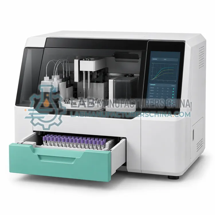
How do Platelet Analyzers work?
Platelet function testing uses two main approaches. In aggregation methods, platelet-rich plasma or whole blood is stirred at 37 degrees Celsius with an agonist such as adenosine diphosphate, collagen, arachidonic acid or ristocetin. In light transmission aggregometry the sample clarifies as platelets clump and the increase in transmitted light is recorded as an aggregation curve. In impedance aggregometry platelets adhere to electrode wires and the rise in electrical impedance between them is recorded. In luminescence aggregometry, ATP released from dense granules reacts with luciferin-luciferase reagent and the emitted light is measured alongside the aggregation trace. In high-shear systems, blood is aspirated through a capillary coated with collagen and adenosine diphosphate or epinephrine, and the time until the aperture occludes is reported as the closure time. All methods depend on controlled temperature, agonist quality and prompt processing of citrated samples.
Which Platelet Analyzers variants are available?
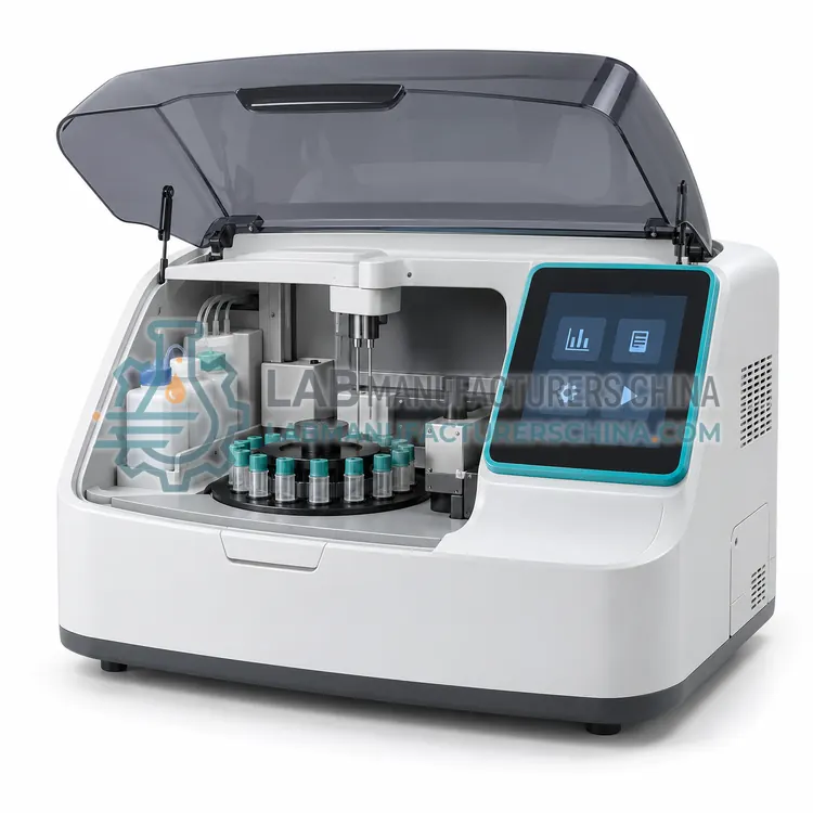
- Optical Platelet Aggregometer — An optical platelet aggregometer measures the increase in light transmitted through stirred platelet-rich plasma as platelets aggregate, producing a curve for each agonist.
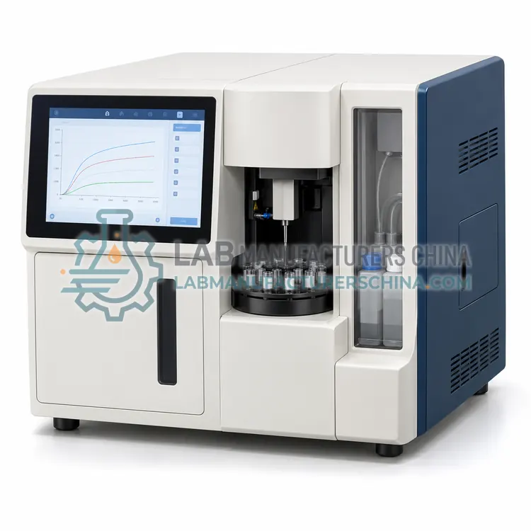
- Whole Blood Impedance Aggregometer — An impedance aggregometer measures the rise in electrical impedance between two electrodes as platelets aggregate onto them in diluted whole blood.
- Lumi-Aggregometer — A lumi-aggregometer measures platelet aggregation and ATP release simultaneously, using a luciferin-luciferase reagent to detect the ATP secreted from dense granules.
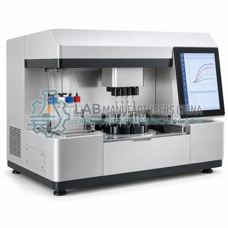
- Multi-Channel Platelet Aggregometer — A multi-channel aggregometer runs four to eight aggregation reactions in parallel, which allows a full agonist panel and controls on one sample at the same time.
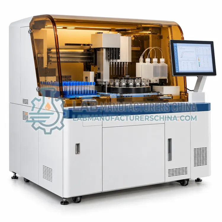
- Automated Platelet Function Analyzer — An automated platelet function analyzer aspirates citrated whole blood through a capillary coated with collagen and an agonist under high shear, and reports the time until the aperture occludes as the closure time.
How do I choose the right variant?
Choose the method according to the clinical question. Light transmission aggregometry is the classical approach and allows a full agonist panel, while impedance aggregometry accepts whole blood and is less affected by low platelet counts and lipemia. Luminescence aggregometry adds dense granule storage pool assessment. High-shear closure time systems are simple to operate and useful for screening and for monitoring antiplatelet therapy, but they give limited mechanistic information. Consider sample volume, which matters for pediatric patients, the number of channels available per run, and the reagent or agonist supply. Confirm whether your laboratory can maintain proficiency in interpretation, because aggregation curves require experience, and take part in external quality assessment where a scheme exists.
Where are Platelet Analyzers used?
Platelet Analyzers are specified in investigation of inherited platelet disorders, testing for aspirin and P2Y12 inhibitor response, diagnosis of von Willebrand disease with ristocetin and uremic and acquired platelet dysfunction assessment.
Which standards and certifications apply?
Work with Platelet Analyzers is commonly governed by ISO 15189, CLSI H58, CLSI EP05, ICSH recommendations on platelet function testing, IEC 61010-1 and IEC 61010-2-101. Instrument certifications such as CE marking are confirmed per configuration and never assumed.
What are the key considerations before purchase?
Collect blood into citrate or hirudin as specified, avoid traumatic venepuncture and process samples within one to four hours, because platelet reactivity declines with time. Keep the aggregometer at 37 degrees Celsius, verify the temperature with an external thermometer and ensure the stir bar rotates at the specified speed. Reagents must be reconstituted and stored exactly as instructed, since degraded agonists give falsely low aggregation. Verify performance with normal control samples and review curves for shape and amplitude, not only the maximum value. Low platelet counts, hemolysis, lipemia and certain drugs affect results, so medication history should be documented before testing.