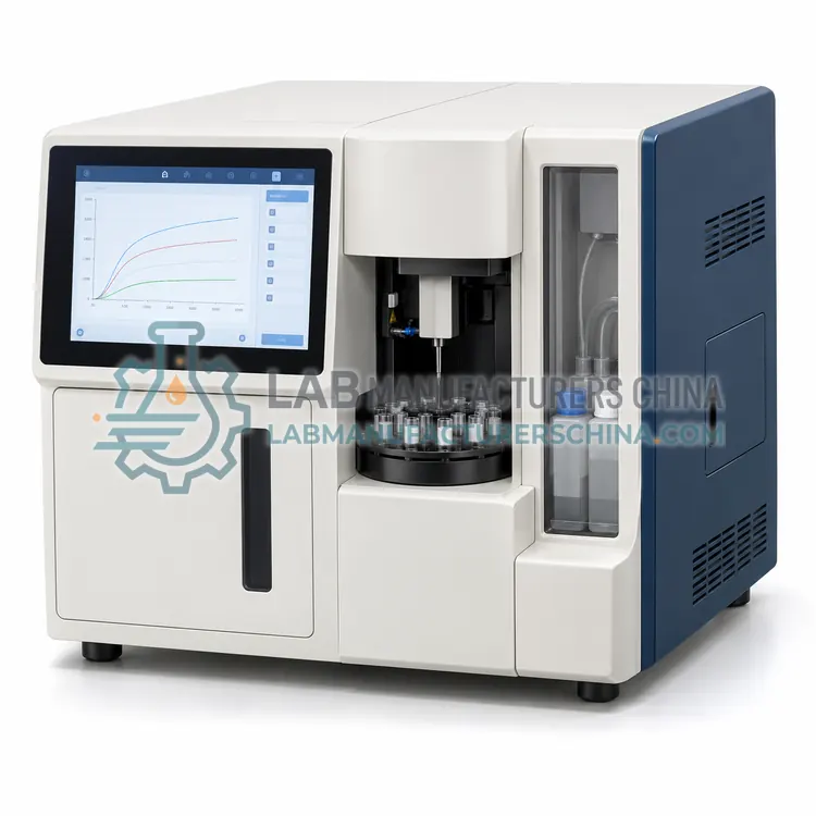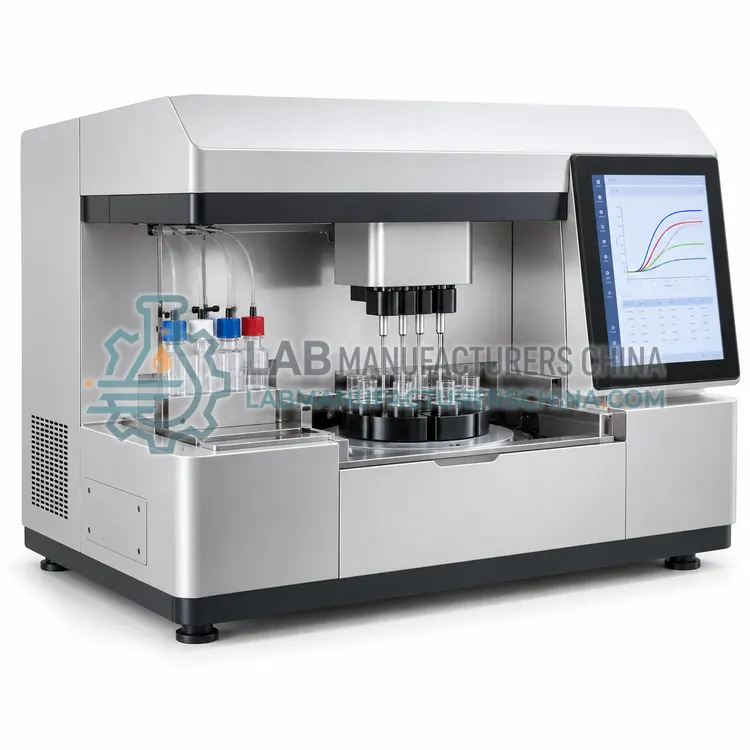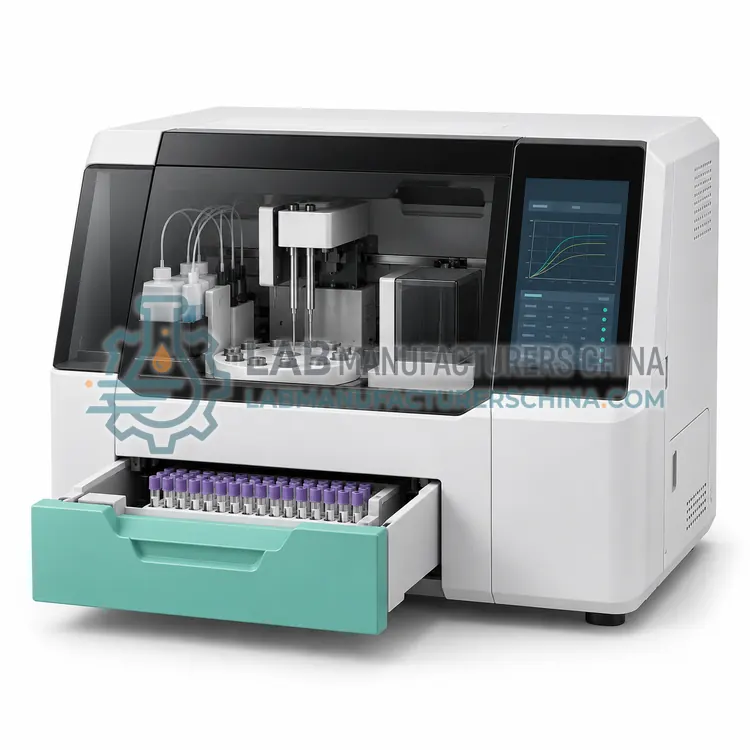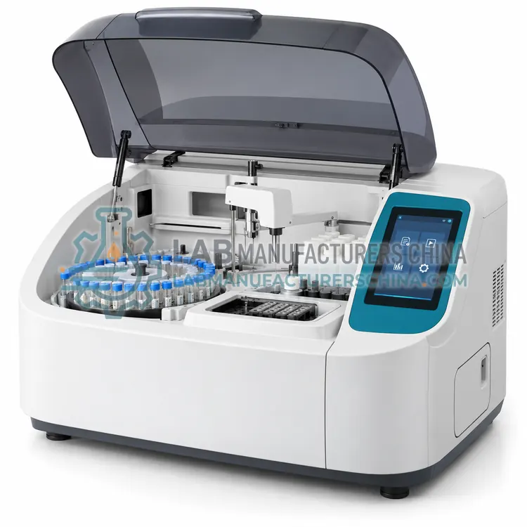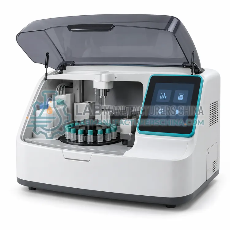
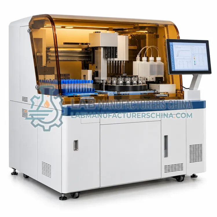
Optical Platelet Aggregometer
光学血小板聚集仪
- SKU PLA-0001
- Platelet Analyzers
- CE (to be confirmed)
- ISO 13485 manufacturing (to be confirmed)
- NMPA class II registration (to be confirmed)
Price on request — FOB / CIF quoted per order
Prices depend on configuration, quantity and destination port.
Key specifications
| Channels | 1–4 cuvette channels |
|---|---|
| Wavelength | 600–650 nm, typically 630 nm |
| Stir speed | 600–1200 rpm adjustable |
| Temperature | 37 °C ±0.2 °C |
| Sample volume | 250–500 µL platelet-rich plasma per channel |
| Agonist volume | 5–50 µL per addition |
Request a quote Download brochure · to be provided
By Lab Manufacturers China Technical TeamLast reviewed Standards: ISO 15189, CLSI H58, CLSI EP05
What is the Optical Platelet Aggregometer?
An optical platelet aggregometer measures the increase in light transmitted through stirred platelet-rich plasma as platelets aggregate, producing a curve for each agonist. It is the classical instrument for diagnosing platelet function disorders and monitoring antiplatelet therapy response.
What is the Optical Platelet Aggregometer?
An optical platelet aggregometer measures the increase in light transmitted through stirred platelet-rich plasma as platelets aggregate, producing a curve for each agonist. It is the classical instrument for diagnosing platelet function disorders and monitoring antiplatelet therapy response.
Overview
The instrument holds one or more cuvette channels at 37 degrees Celsius, each with a light source, a photodetector and a magnetic stirrer. Platelet-rich plasma is prepared by low-speed centrifugation, an agonist is added after a stable baseline is recorded, and the change in light transmission is plotted against time. Software derives the maximum aggregation percentage, the slope, the lag time and, with a second phase, the disaggregation pattern. Because the method requires platelet-rich plasma, the platelet count must be adjusted within a defined range for results to be comparable with reference patterns, and lipemic or hemolyzed samples are unsuitable. Interpretation depends on comparing the response profile to several agonists, which requires trained staff and clearly documented reference patterns.
How does it work?
Platelet function testing uses two main approaches. In aggregation methods, platelet-rich plasma or whole blood is stirred at 37 degrees Celsius with an agonist such as adenosine diphosphate, collagen, arachidonic acid or ristocetin. In light transmission aggregometry the sample clarifies as platelets clump and the increase in transmitted light is recorded as an aggregation curve. In impedance aggregometry platelets adhere to electrode wires and the rise in electrical impedance between them is recorded. In luminescence aggregometry, ATP released from dense granules reacts with luciferin-luciferase reagent and the emitted light is measured alongside the aggregation trace. In high-shear systems, blood is aspirated through a capillary coated with collagen and adenosine diphosphate or epinephrine, and the time until the aperture occludes is reported as the closure time. All methods depend on controlled temperature, agonist quality and prompt processing of citrated samples.
What are the key features?
- One to four cuvette channels with individual stirrers at 37 °C
- Light transmission measurement against a plasma blank
- Software calculation of maximum aggregation, slope and lag time
- Second-phase disaggregation detection in some models
- Adjustable stir speed and baseline stabilization time
- Curve storage for comparative interpretation
What are the technical specifications?
A typical optical platelet aggregometer configuration offers channels of 1–4 cuvette channels, wavelength of 600–650 nm, typically 630 nm, stir speed of 600–1200 rpm adjustable, temperature of 37 °C ±0.2 °C, sample volume of 250–500 µL platelet-rich plasma per channel and agonist volume of 5–50 µL per addition. Values are indicative of the product class; the exact figures for each configuration are confirmed on the quotation and the calibration documents supplied with the instrument.
Which applications is it used for?
The Optical Platelet Aggregometer is used in inherited platelet disorder diagnosis, aspirin and P2Y12 inhibitor response testing, von Willebrand disease investigation with ristocetin and pre-operative platelet function assessment. Across the wider Platelet Analyzers class, laboratories also deploy it for investigation of inherited platelet disorders, testing for aspirin and P2Y12 inhibitor response and diagnosis of von Willebrand disease with ristocetin.
Which standards and certifications apply?
Relevant standards and methods include ISO 15189, CLSI H58, CLSI EP05, ICSH recommendations on platelet function testing, IEC 61010-1 and IEC 61010-2-101. Certification candidates for this model are CE, ISO 13485 manufacturing and NMPA class II registration; each is to be confirmed for the specific configuration before shipment, and certificates are supplied only where they have been issued.
Collect blood into citrate or hirudin as specified, avoid traumatic venepuncture and process samples within one to four hours, because platelet reactivity declines with time. Keep the aggregometer at 37 degrees Celsius, verify the temperature with an external thermometer and ensure the stir bar rotates at the specified speed. Reagents must be reconstituted and stored exactly as instructed, since degraded agonists give falsely low aggregation. Verify performance with normal control samples and review curves for shape and amplitude, not only the maximum value. Low platelet counts, hemolysis, lipemia and certain drugs affect results, so medication history should be documented before testing.
How do I select the right Optical Platelet Aggregometer?
Specify this variant when a full agonist panel is needed to characterize a suspected platelet defect, or when the laboratory requires the classical reference approach. In the RFQ confirm the number of channels, temperature stability, stir speed range and software curve analysis, and plan the sample preparation workload, since platelet-rich plasma must be prepared and counted before each run.
How does it compare with other Platelet Analyzers variants?
Choose the method according to the clinical question. Light transmission aggregometry is the classical approach and allows a full agonist panel, while impedance aggregometry accepts whole blood and is less affected by low platelet counts and lipemia. Luminescence aggregometry adds dense granule storage pool assessment. High-shear closure time systems are simple to operate and useful for screening and for monitoring antiplatelet therapy, but they give limited mechanistic information. Consider sample volume, which matters for pediatric patients, the number of channels available per run, and the reagent or agonist supply. Confirm whether your laboratory can maintain proficiency in interpretation, because aggregation curves require experience, and take part in external quality assessment where a scheme exists.
Full specifications
| Channels | 1–4 cuvette channels |
|---|---|
| Wavelength | 600–650 nm, typically 630 nm |
| Stir speed | 600–1200 rpm adjustable |
| Temperature | 37 °C ±0.2 °C |
| Sample volume | 250–500 µL platelet-rich plasma per channel |
| Agonist volume | 5–50 µL per addition |
| Parameters reported | Maximum aggregation %, slope, lag time, disaggregation |
| Data storage | 500 curves with test identifiers |
| Interfaces | USB, RS-232 with software export |
Indicative values for this product class. Final specifications are confirmed on the quotation and in the calibration documents supplied with the instrument.
Applications
- Inherited platelet disorder diagnosis
- Aspirin and P2Y12 inhibitor response testing
- Von Willebrand disease investigation with ristocetin
- Pre-operative platelet function assessment
Key features
- One to four cuvette channels with individual stirrers at 37 °C
- Light transmission measurement against a plasma blank
- Software calculation of maximum aggregation, slope and lag time
- Second-phase disaggregation detection in some models
- Adjustable stir speed and baseline stabilization time
- Curve storage for comparative interpretation
Frequently asked questions about Optical Platelet Aggregometer
Blood is centrifuged at low speed, typically 150 to 200 times gravity for ten to fifteen minutes, and the supernatant plasma is collected. The platelet count is then adjusted with platelet-poor plasma to a defined range, usually 200 to 250 ×10⁹/L, so that curves can be compared with reference patterns.
Yes, because the method relies on changes in light transmission, and lipids or free hemoglobin reduce the optical clarity of the plasma. Such samples should be reported with a comment or the test repeated when the patient's condition allows, and impedance aggregometry is an alternative for whole blood samples.
A normal response shows a small initial shape-change phase, then a rapid rise to a stable or slightly reversible plateau, with the maximum percentage depending on the agonist and its concentration. Laboratories should establish their own reference patterns using normal donor samples.
Standard light transmission aggregometers require platelet-rich plasma, so whole blood cannot be used directly. Laboratories that prefer whole blood should choose an impedance aggregometer, which also tolerates low platelet counts and lipemic samples better.
It measures the increase in light transmitted through stirred platelet-rich plasma as platelets aggregate in response to an agonist. The resulting curve shows the rate, extent and reversibility of aggregation for each agonist, and comparison with normal patterns supports the diagnosis of specific platelet disorders.
Common agonists are adenosine diphosphate to assess P2Y12 and P2Y1 receptors, arachidonic acid to assess the cyclooxygenase pathway, collagen for the glycoprotein VI pathway, ristocetin for von Willebrand factor and glycoprotein Ib, and epinephrine or thrombin receptor peptide for additional receptor studies.
Collect blood with minimal stasis into citrate or, for some assays, hirudin, keep it at room temperature and test within one to four hours. Do not refrigerate, shake vigorously or delay processing, because platelet activation and disaggregation change the curves and can make interpretation unreliable.
Related models