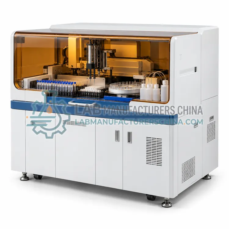
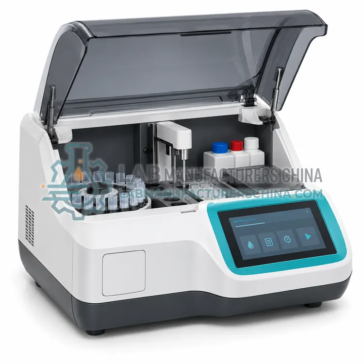
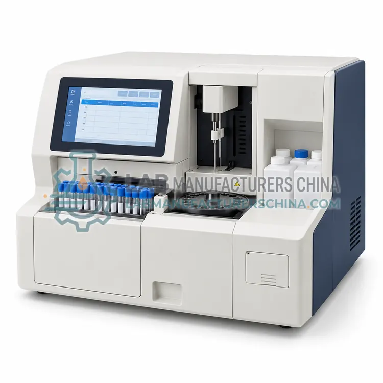
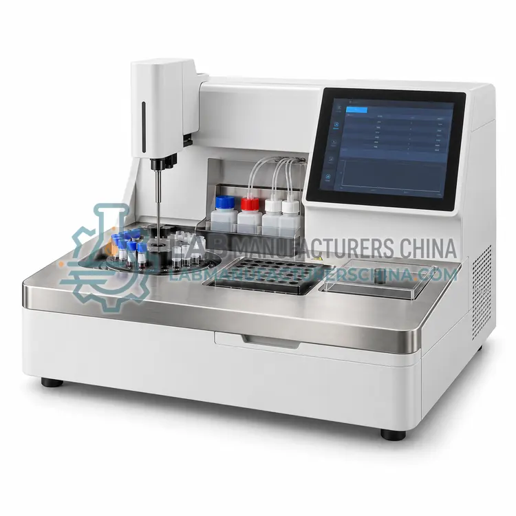
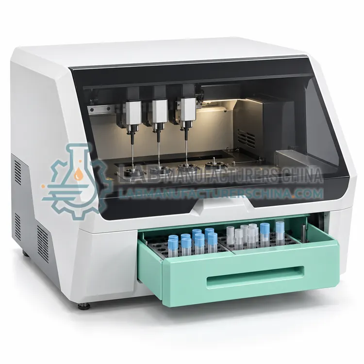
How do Semi-Automatic Coagulation Analyzers work?
A semi-automatic coagulation analyzer provides a temperature-controlled measuring position, usually at 37 degrees Celsius, a detection system and a timer. The operator pipettes plasma into the cuvette, incubates it, adds the reagent and starts the measurement. Optical instruments detect the increase in turbidity or the change in light scatter as fibrin strands form; mechanical instruments detect the change in movement or viscosity of a steel ball or magnetic particle; turbidimetric and chromogenic instruments measure light absorption changes from the fibrin network or from a coloured product released by a synthetic substrate. The analyzer stops the timer at the detection threshold and reports the clotting time in seconds, a ratio for prothrombin time or a concentration for fibrinogen. Because sample and reagent handling remain manual, pipetting accuracy and consistent incubation timing have a large influence on results.
Which Semi-Automatic Coagulation Analyzers variants are available?
- Semi-Automatic Coagulation Analyzer with Optical Clot Detection — This analyzer detects clot formation optically by measuring the change in light scatter as fibrin forms in the cuvette.
- Semi-Automatic Coagulation Analyzer with Mechanical Clot Detection — This analyzer detects clot formation mechanically by monitoring the movement of a steel ball or magnetic particle as plasma viscosity rises.
- Semi-Automatic Coagulation Analyzer with Turbidimetric Detection — This analyzer measures the increase in turbidity as fibrinogen is converted to fibrin, using a photometric channel that is well suited to fibrinogen assays and to reagents with a strong optical signal.
- Semi-Automatic Coagulation Analyzer with Chromogenic Capability — This analyzer adds a chromogenic channel that measures colour released from a synthetic substrate by a coagulation enzyme or inhibitor, enabling assays such as antithrombin and protein C.
- Four-Channel Semi-Automatic Coagulation Analyzer — This instrument provides four independent measuring channels so that a patient sample, a control and duplicates can run in parallel, reducing operator waiting time.
How do I choose the right variant?
Choose the detection principle according to the samples you receive: mechanical or magnetic detection tolerates icteric, hemolyzed and lipemic plasma, while optical detection is simpler and usually requires a clear sample. Then decide how many channels you need, because a four-channel instrument allows several patients or duplicate measurements in one run and reduces operator waiting time. Check the menu the instrument supports, especially whether fibrinogen, D-dimer or antithrombin assays are available, since not every semi-automatic platform can perform chromogenic or immunoturbidimetric tests. Confirm the sample and reagent volumes, the incubation temperature accuracy and the calibration requirements, and ask whether the analyzer stores calibrator values for each reagent lot.
Where are Semi-Automatic Coagulation Analyzers used?
Semi-Automatic Coagulation Analyzers are specified in routine PT and APTT testing in clinics, anticoagulation monitoring with INR, pre-operative coagulation screening and fibrinogen measurement in small laboratories.
Which standards and certifications apply?
Work with Semi-Automatic Coagulation Analyzers is commonly governed by ISO 15189, CLSI H57, CLSI H21, CLSI EP05, ICSH recommendations on coagulation testing, IEC 61010-1 and IEC 61010-2-101. Instrument certifications such as CE marking are confirmed per configuration and never assumed.
What are the key considerations before purchase?
Place the analyzer on a level bench away from vibration and draughts, since both disturb optical clot detection, and allow the instrument to reach and hold 37 degrees Celsius before use. Verify performance at installation against a reference instrument or assigned plasma samples for prothrombin time, APTT and fibrinogen, and check the incubation temperature with an external thermometer. Daily work includes running normal and abnormal control plasma, checking reagent and cuvette lot numbers and recording all values. Reagents must be reconstituted, stored and expired strictly according to the instructions, and citrated plasma should be separated, frozen and thawed under controlled conditions because pre-analytical handling has a large influence on clotting times.