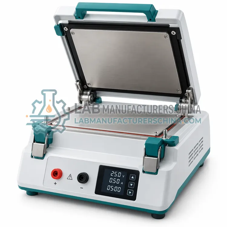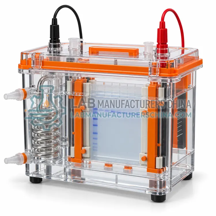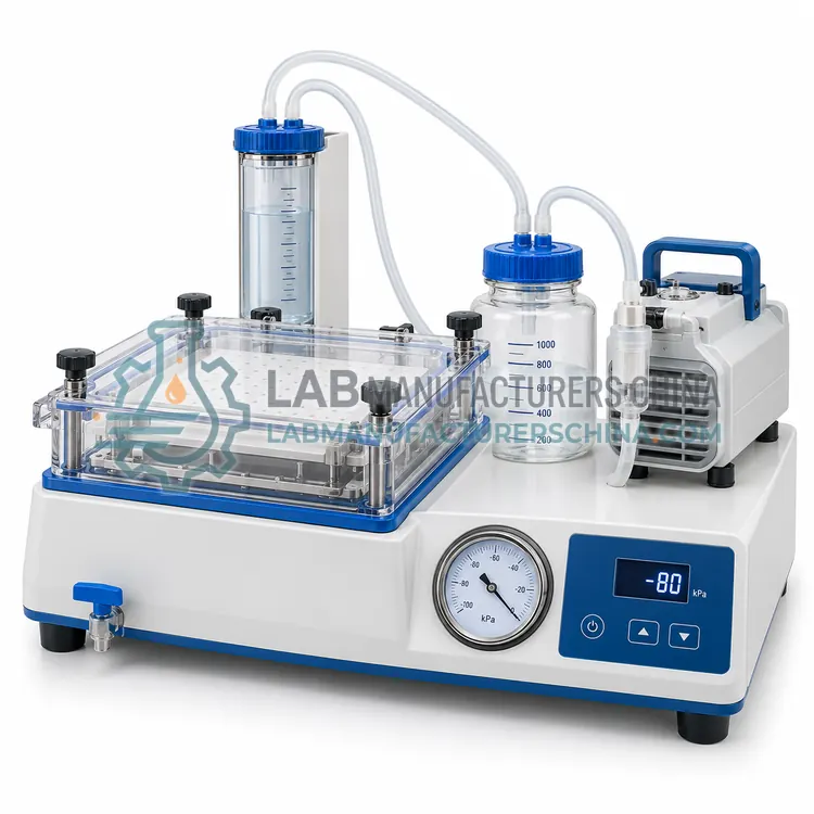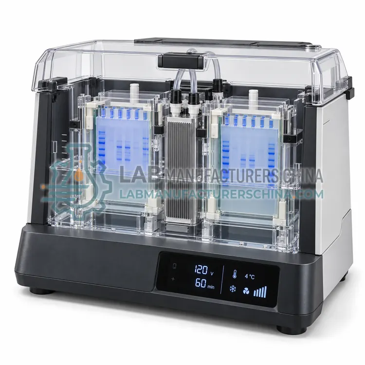
Automated Western Blot Incubation System
自动化 Western 印迹孵育系统
- SKU WBS-0005
- Western Blot Systems
- CE (to be confirmed)
- ISO 9001 manufacturing (to be confirmed)
- IEC 61010-1 safety (to be confirmed)
Price on request — FOB / CIF quoted per order
Prices depend on configuration, quantity and destination port.
Key specifications
| Membranes per run | 1–6 (model dependent) |
|---|---|
| Antibody volume | 1–5 mL per membrane |
| Incubation time range | 1 min to overnight |
| Temperature control | ambient to 40 °C |
| Agitation | Rocking or orbital, 5–60 rpm |
| Wash steps | Programmable, up to 10 repetitions |
Request a quote Download brochure · to be provided
By Lab Manufacturers China Technical TeamLast reviewed Standards: USP <1057>, Ph. Eur. 2.2.31, IEC 61010-1
What is the Automated Western Blot Incubation System?
An automated incubation system performs blocking, antibody incubation and washing on membranes in a programmable tray or cassette, replacing manual shaking trays, reducing antibody consumption and improving consistency between blots in routine laboratories.
What is the Automated Western Blot Incubation System?
An automated incubation system performs blocking, antibody incubation and washing on membranes in a programmable tray or cassette, replacing manual shaking trays, reducing antibody consumption and improving consistency between blots in routine laboratories.
Overview
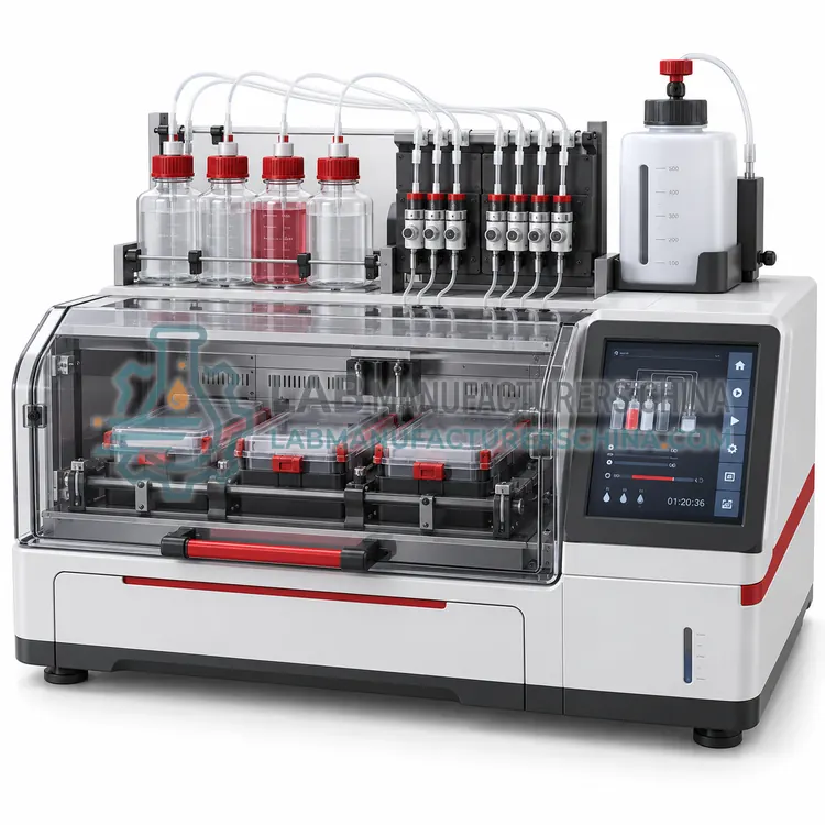
The system holds one or several membranes in individual compartments and delivers blocking buffer, antibody solutions and wash buffer by programmable pumps, with rocking or orbital agitation and controlled temperature. Volumes are typically 1–5 mL per membrane, considerably less than in manual trays, which lowers antibody cost for expensive reagents. Programs define incubation times from minutes to overnight, number of washes and temperature, and several membranes can be processed with different antibodies in the same run. Because timing and volume are instrument controlled, background and signal intensity are more consistent between blots, which matters for comparative quantification. Instruments usually include reagent recovery, waste collection and a log of protocol steps for traceability, and some models handle gel staining and membrane stripping in the same run.
How does it work?
Western blotting starts with separation of proteins by SDS-PAGE or a capillary system. In conventional blotting, the gel is placed against a nitrocellulose or PVDF membrane in a transfer stack and an electric field drives the negatively charged proteins out of the gel onto the membrane, where they bind by hydrophobic and electrostatic interaction. The membrane is then blocked with protein or detergent to saturate remaining binding sites, incubated with a primary antibody against the target, washed, incubated with an enzyme- or fluorophore-labelled secondary antibody that recognises the primary antibody, and washed again. Bound enzyme is detected by adding a chemiluminescent or colorimetric substrate, and the signal is imaged and quantified against a standard or a loading control. Automated capillary systems perform the equivalent steps in a capillary with a separation matrix and immobilised reagents.
What are the key features?
- Programmable delivery of blocking, antibody and wash solutions
- Individual membrane compartments to prevent cross-contamination
- Antibody volumes of 1–5 mL per membrane
- Controlled rocking or orbital agitation with temperature option
- Protocol logging and reagent recovery functions
- Automated stripping and re-probing of the same membrane
What are the technical specifications?
A typical automated western blot incubation system configuration offers membranes per run of 1–6 (model dependent), antibody volume of 1–5 mL per membrane, incubation time range of 1 min to overnight, temperature control of ambient to 40 °C, agitation of Rocking or orbital, 5–60 rpm and wash steps of Programmable, up to 10 repetitions. Values are indicative of the product class; the exact figures for each configuration are confirmed on the quotation and the calibration documents supplied with the instrument.
Which applications is it used for?
The Automated Western Blot Incubation System is used in repetitive blot processing in quality control, multi-antibody screening on replicate membranes, phosphoprotein analysis with sensitive antibodies and long overnight incubations without manual attendance. Across the wider Western Blot Systems class, laboratories also deploy it for specific protein detection in cell lysates, recombinant protein expression monitoring and biopharmaceutical purity and identity testing.
Which standards and certifications apply?
Relevant standards and methods include USP <1057>, Ph. Eur. 2.2.31, IEC 61010-1, ISO 17025, 21 CFR Part 11 and ISO 9001. Certification candidates for this model are CE, ISO 9001 manufacturing and IEC 61010-1 safety; each is to be confirmed for the specific configuration before shipment, and certificates are supplied only where they have been issued.
Transfer efficiency depends on current, time, buffer composition and the amount of methanol or SDS, so conditions should be established for each target protein and recorded in the method. Air bubbles trapped in the stack cause localised transfer gaps, and membranes must be handled with gloves and kept wet once activated. Blocking agent choice affects background and antibody binding, and milk-based blockers can interfere with phospho-specific antibodies. Wash volumes and times must be consistent to keep background low. Chemiluminescent substrates fade quickly, so images should be captured within the recommended window, and quantitative comparisons should include a loading control on the same membrane. Store antibodies in aliquots at the recommended temperature and record lot numbers, because antibody performance varies between lots.
How do I select the right Automated Western Blot Incubation System?
Select an automated incubation system when antibody cost, incubation consistency or unattended overnight processing matters, or when several membranes must be treated identically. Specify membrane capacity, antibody volume, temperature range, agitation control, reagent recovery and protocol logging.
How does it compare with other Western Blot Systems variants?
Choose first between conventional and automated blotting. Conventional tank or semi-dry transfer with manual antibody incubation costs less per sample and allows flexible antibody choices, but requires more hands-on time and gives semi-quantitative results. Automated capillary systems reduce run time to a few hours, use nanolitre sample volumes and produce quantitative peak areas, which suits regulated laboratories generating many comparable data sets. For conventional workflows, decide transfer format by gel size and protein molecular weight: tank transfer suits large gels and high molecular weight proteins, semi-dry transfer is faster and uses less buffer, and vacuum transfer is useful for thick gels. Check membrane and gel dimensions, electrode type, current and voltage range, cooling options and whether the system handles multiple gels at once.
Full specifications
| Membranes per run | 1–6 (model dependent) |
|---|---|
| Antibody volume | 1–5 mL per membrane |
| Incubation time range | 1 min to overnight |
| Temperature control | ambient to 40 °C |
| Agitation | Rocking or orbital, 5–60 rpm |
| Wash steps | Programmable, up to 10 repetitions |
| Reagent recovery | Antibody solutions recoverable for reuse |
| Protocol storage | Multiple programs with step logging |
| Membrane formats | Mini and midi blot sizes |
| Dimensions (W x D x H) | approx. 400 x 350 x 250 mm |
Indicative values for this product class. Final specifications are confirmed on the quotation and in the calibration documents supplied with the instrument.
Applications
- Repetitive blot processing in quality control
- Multi-antibody screening on replicate membranes
- Phosphoprotein analysis with sensitive antibodies
- Long overnight incubations without manual attendance
Key features
- Programmable delivery of blocking, antibody and wash solutions
- Individual membrane compartments to prevent cross-contamination
- Antibody volumes of 1–5 mL per membrane
- Controlled rocking or orbital agitation with temperature option
- Protocol logging and reagent recovery functions
- Automated stripping and re-probing of the same membrane
Frequently asked questions about Automated Western Blot Incubation System
Typical working volumes are 1–5 mL per membrane instead of 10–20 mL in a manual tray, so antibody consumption can fall by 60–90%. Recovered primary antibody solutions can often be reused two or three times if stored correctly.
Yes, when each membrane has its own compartment and reagent line. Programs can assign separate antibody solutions and incubation times to each compartment, which is useful for screening several targets in one run.
Signal intensity is usually similar to manual processing, but consistency improves because timing, volume and agitation are fixed. Laboratories should still compare results with their established manual method and set acceptance criteria during validation.
An electric field drives negatively charged SDS-coated proteins out of the gel and onto a nitrocellulose or PVDF membrane placed against it. Proteins bind the membrane by hydrophobic and electrostatic interactions, after which antibodies detect the target.
Nitrocellulose binds proteins well and gives low background for most chemiluminescent detection. PVDF has higher binding capacity and better mechanical strength, which helps with repeated stripping and reprobing and with low-abundance or small proteins.
Use a pre-stained molecular weight ladder on the gel, and stain the membrane with Ponceau S or a reversible protein stain to confirm that proteins have moved. Staining the gel after transfer shows how much protein remains behind.
Related models