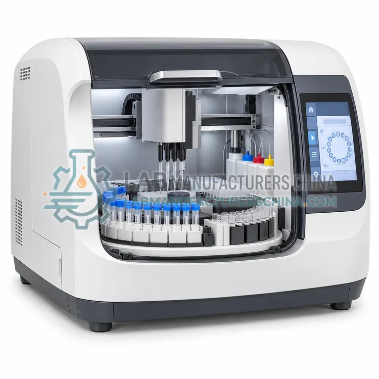
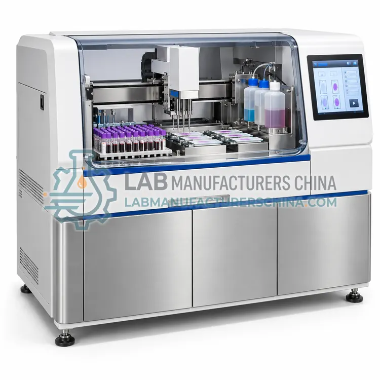
Automated Blood Smear Preparation System
自动血涂片制备系统
- SKU SAM-0005
- Sample Preparation
- CE (to be confirmed)
- ISO 13485 manufacturing (to be confirmed)
- NMPA class II registration (to be confirmed)
Price on request — FOB / CIF quoted per order
Prices depend on configuration, quantity and destination port.
Key specifications
| Sample volume | 2–10 µL blood per slide |
|---|---|
| Spread parameters | Adjustable angle, drop volume and speed |
| Hematocrit adjustment | Preset profiles for low, normal and high Hct |
| Stain | Romanowsky-type stain, onboard bottles |
| Throughput | 30–60 slides/h |
| Slide identification | Printed identifier or barcode label |
Request a quote Download brochure · to be provided
By Lab Manufacturers China Technical TeamLast reviewed Standards: ISO 15189, ISO 15190, CLSI GP16
What is the Automated Blood Smear Preparation System?
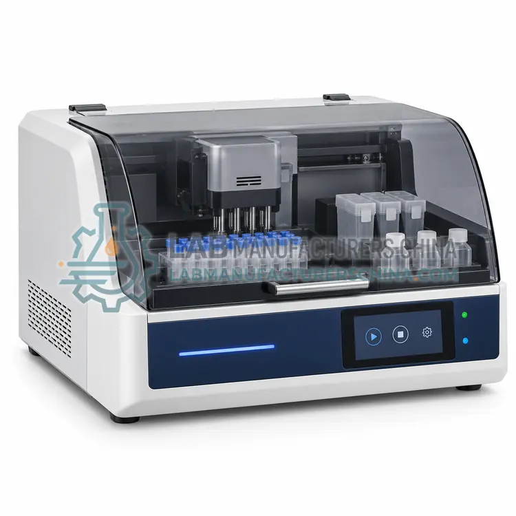
An automated blood smear preparation system aspirates blood, spreads a monolayer slide and stains it with a Romanowsky stain at defined settings. It suits hematology workflows that need consistent films for morphological review, including veterinary and research laboratories.
What is the Automated Blood Smear Preparation System?
An automated blood smear preparation system aspirates blood, spreads a monolayer slide and stains it with a Romanowsky stain at defined settings. It suits hematology workflows that need consistent films for morphological review, including veterinary and research laboratories.
Overview
The device takes a small volume of EDTA or capillary blood, deposits it on a slide and draws a spreader at a controlled angle and speed so that a monolayer forms where cells are evenly distributed and not overlapping. Slides are dried and stained with a Romanowsky stain, and the instrument prints or applies an identifier. Spread angle, drop size and push speed are adjusted for hematocrit, because viscous samples from anaemic or polycythaemic patients need different settings to achieve a readable monolayer. Some systems integrate with a cell counter so that slide preparation can be triggered automatically by review flags. Quality is assessed by monolayer length, even distribution and correct staining, and slides should be inspected during validation and in periodic quality checks.
How does it work?
Diagnostic sample preparation changes a specimen so that a downstream assay can measure it reliably. Serum and plasma are separated from cells by centrifugation and then aliquoted into labelled tubes, which reduces thaw cycles and cross-contamination during archiving. Some serology and molecular workflows require heat inactivation, typically at 56 degrees Celsius for thirty minutes, to reduce infectivity before handling. Solid or viscous specimens such as stool, sputum, swabs and tissue must be homogenised, liquefied or extracted into a buffer so that target organisms or analytes are released uniformly. Fecal immunochemical tests need a defined sample mass mixed with buffer, and blood film preparation requires a controlled spread and stain so that cells can be examined morphologically. Each step influences the analytical result, so volume accuracy, temperature control and traceability matter as much as in the analytical instrument itself.
What are the key features?
- Controlled spread angle and speed for consistent monolayers
- Hematocrit-based adjustment of drop size and push speed
- Romanowsky staining with onboard stain bottles
- Printed or barcoded slide identification
- Optional triggering by cell counter review flags
- Slide quality assessment by monolayer length and distribution
What are the technical specifications?
A typical automated blood smear preparation system configuration offers sample volume of 2–10 µL blood per slide, spread parameters of Adjustable angle, drop volume and speed, hematocrit adjustment of Preset profiles for low, normal and high Hct, stain of Romanowsky-type stain, onboard bottles, throughput of 30–60 slides/h and slide identification of Printed identifier or barcode label. Values are indicative of the product class; the exact figures for each configuration are confirmed on the quotation and the calibration documents supplied with the instrument.
Which applications is it used for?
The Automated Blood Smear Preparation System is used in hematology blood film preparation for review, veterinary blood smear preparation, research blood film work and teaching laboratories demonstrating cell morphology. Across the wider Sample Preparation class, laboratories also deploy it for serum and plasma aliquotting for serology and molecular testing, heat inactivation of serum before batch serology and stool and sputum processing for infectious disease testing.
Which standards and certifications apply?
Relevant standards and methods include ISO 15189, ISO 15190, CLSI GP16, CLSI M29, CLSI EP05, IEC 61010-1 and IEC 61010-2-101. Certification candidates for this model are CE, ISO 13485 manufacturing and NMPA class II registration; each is to be confirmed for the specific configuration before shipment, and certificates are supplied only where they have been issued.
Verify each device at installation with gravimetric volume checks, temperature mapping and, where relevant, recovery or uniformity studies using spiked specimens. Write procedures that define specimen acceptance, batch size, temperature and time limits, and the identification of each aliquot, because traceability between the primary tube and derived aliquots is frequently examined during accreditation. Clean and decontaminate surfaces, rotors and homogenisation chambers after each batch, and use sealed or disposable components where aerosols are a risk. Record maintenance, calibration and validation data, and review temperature records for heat inactivation and incubation steps. Treat all specimens, consumables and waste as potentially infectious.
How do I select the right Automated Blood Smear Preparation System?
Specify this system when blood films are needed consistently, when manual spreading produces variable quality, or when slide preparation should be triggered automatically by analyzer flags. In the RFQ confirm hematocrit adjustment options, stain type and consumption, slide capacity and whether barcode identification can be linked to the analyzer record.
How does it compare with other Sample Preparation variants?
Start from the specimen types you receive and the assays that follow, since a device is only useful if its process matches the downstream kit requirements. For aliquotting, check the number of tubes per primary sample, the dead volume, the liquid class accuracy for viscous specimens and the labelling method. For heat inactivation, confirm the temperature uniformity across the bath or block, the capacity per batch and whether a lid is needed to limit evaporation. For homogenisation, choose between bead milling, rotor-stator and paddle systems according to specimen hardness and the need to keep analytes intact. For fecal and blood smear preparation, confirm the sample mass or film quality specification. In all cases, ask for process validation data and confirm that consumables are available locally.
Full specifications
| Sample volume | 2–10 µL blood per slide |
|---|---|
| Spread parameters | Adjustable angle, drop volume and speed |
| Hematocrit adjustment | Preset profiles for low, normal and high Hct |
| Stain | Romanowsky-type stain, onboard bottles |
| Throughput | 30–60 slides/h |
| Slide identification | Printed identifier or barcode label |
| Stain consumption | About 1 mL per slide |
| Data records | Run log with slide identification |
| Power supply | AC 100–240 V, 50/60 Hz |
Indicative values for this product class. Final specifications are confirmed on the quotation and in the calibration documents supplied with the instrument.
Applications
- Hematology blood film preparation for review
- Veterinary blood smear preparation
- Research blood film work
- Teaching laboratories demonstrating cell morphology
Key features
- Controlled spread angle and speed for consistent monolayers
- Hematocrit-based adjustment of drop size and push speed
- Romanowsky staining with onboard stain bottles
- Printed or barcoded slide identification
- Optional triggering by cell counter review flags
- Slide quality assessment by monolayer length and distribution
Frequently asked questions about Automated Blood Smear Preparation System
High hematocrit blood is more viscous and spreads into a shorter, thicker film, while low hematocrit blood spreads thinly and may extend beyond the readable area. The instrument adjusts drop size, angle or speed, or the laboratory dilutes the sample, to obtain a monolayer suitable for examination.
Yes, films prepared with reproducible spread and stain are suitable for digital morphology scanners, which locate the monolayer automatically. Consistent preparation improves the reliability of image analysis and reduces the number of slides that must be repeated.
Check that the monolayer is of adequate length, that cells are evenly distributed without overlap or clumping, and that staining shows correct nuclear and cytoplasmic colours. Inspect slides during validation and at defined intervals, and record the findings with corrective actions where needed.
Yes, it uses Romanowsky-type stain components that must be stored and replaced per the manufacturer schedule, typically consuming about one millilitre per slide. Stain deterioration changes colour balance, so replacement intervals should be based on volume and time, not only on visible discolouration.
Heat inactivation at 56 degrees Celsius for about thirty minutes reduces the infectivity of serum and plasma before they are handled in batches or outside a biosafety cabinet, which lowers the risk to staff. It must be validated for each assay, because heating can degrade some analytes and enzymes.
It depends on the primary tube volume, the dead volume of the pipetting system, and the volume required per test. Laboratories usually plan two to four aliquots per serum tube to limit thaw cycles, and the pipetting station should be specified with the minimum deliverable volume that remains accurate.
Bead milling with stainless steel or ceramic beads suits fibrous tissue and tough specimens, while rotor-stator homogenisers handle larger volumes of sputum and tissue quickly. Chemical liquefaction with a reducing agent such as dithiothreitol is often combined with mechanical processing for sputum in mycobacteriology.
Related models