
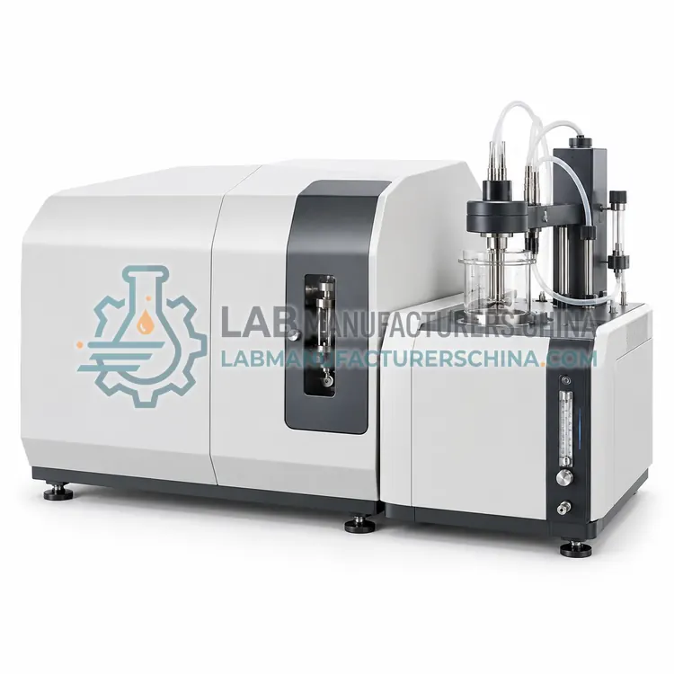
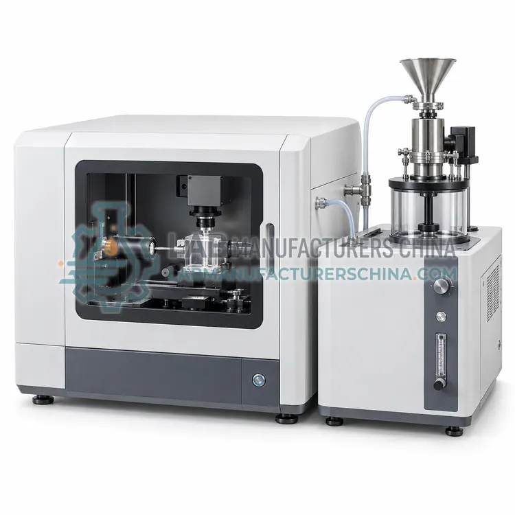
Dynamic Image Particle Size and Shape Analyzer
动态图像粒度粒形分析仪
- SKU PSA-0004
- Particle Size Analyzers
- CE (to be confirmed)
- ISO 9001 manufacturing (to be confirmed)
Price on request — FOB / CIF quoted per order
Prices depend on configuration, quantity and destination port.
Key specifications
| Measuring range | 1 µm–30 mm (optics dependent) |
|---|---|
| Measurement principle | Dynamic image analysis (ISO 13322-2) |
| Cameras | Two, up to 300 fps |
| Light source | Pulsed LED backlight |
| Dispersion modes | Free fall, air, wet cell |
| Size parameters | xc min, xFe max, area equivalent |
Request a quote Download brochure · to be provided
By Lab Manufacturers China Technical TeamLast reviewed Standards: ISO 13320, USP <429>, ISO 13322-1
What is the Dynamic Image Particle Size and Shape Analyzer?
An image-based analyzer that films particles in free fall or flow with high-speed cameras and reports size together with shape parameters such as aspect ratio and circularity. It is chosen when particle morphology matters or when rare oversize particles must be detected.
What is the Dynamic Image Particle Size and Shape Analyzer?
An image-based analyzer that films particles in free fall or flow with high-speed cameras and reports size together with shape parameters such as aspect ratio and circularity. It is chosen when particle morphology matters or when rare oversize particles must be detected.
Overview
Particles are dispersed by a vibratory feeder and gravity, compressed air or a liquid flow cell and imaged against a pulsed LED backlight by one or two cameras at high frame rates, often with zoom and basic optics for different size windows. Software extracts each particle silhouette and calculates equivalent diameters such as area-equivalent, Feret maximum and minimum, plus aspect ratio, convexity and circularity, as described in ISO 13322-2. Because every particle is counted individually, the method resolves a few percent of oversize or agglomerated particles that laser diffraction averages out. Typical ranges are 1 µm to 30 mm depending on optics. Hundreds of thousands of particles per minute give good statistics, and stored images provide traceable evidence for investigations.
How does it work?
Laser diffraction analyzers pass a laser beam through a dispersed sample; particles scatter light at angles inversely related to their size, and a detector array records the angular intensity pattern. The Mie or Fraunhofer model converts this pattern into a volume-based size distribution. Dynamic light scattering instead measures intensity fluctuations caused by Brownian motion, and the Stokes–Einstein equation converts the diffusion coefficient into a hydrodynamic diameter. Dynamic image analysis captures particle silhouettes with a high-speed camera and reports size and shape descriptors directly. Sedimentation methods apply Stokes' law to settling velocity under gravity or centrifugal force. Key components include the light source, sample dispersion unit (wet or dry), detector array or correlator, and software with optical-property databases.
What are the key features?
- Dual-camera zoom and basic optics for wide dynamic range
- Pulsed LED backlight to freeze fast-moving particles
- Size and shape parameters including aspect ratio and circularity
- Dry free-fall, air-dispersion and wet flow-cell modules
- Stored particle images for audit and investigation
- Detection of oversize or agglomerated particles at low percentages
What are the technical specifications?
A typical dynamic image particle size and shape analyzer configuration offers measuring range of 1 µm–30 mm (optics dependent), measurement principle of Dynamic image analysis (ISO 13322-2), cameras of Two, up to 300 fps, light source of Pulsed LED backlight, dispersion modes of Free fall, air, wet cell and size parameters of xc min, xFe max, area equivalent. Values are indicative of the product class; the exact figures for each configuration are confirmed on the quotation and the calibration documents supplied with the instrument.
Which applications is it used for?
The Dynamic Image Particle Size and Shape Analyzer is used in abrasives, proppants and glass beads, metal powders for additive manufacturing, fertiliser and granulate shape control, crystalline API morphology screening and food ingredients and salt. Across the wider Particle Size Analyzers class, laboratories also deploy it for pharmaceutical powder and inhaler development, battery cathode and anode materials and ceramics and cement.
Which standards and certifications apply?
Relevant standards and methods include ISO 13320, USP <429>, ISO 13322-1, ISO 13322-2, ISO 22412, GB/T 19077 and Ph. Eur. 2.9.31. Certification candidates for this model are CE and ISO 9001 manufacturing; each is to be confirmed for the specific configuration before shipment, and certificates are supplied only where they have been issued.
Particle size results depend heavily on sampling and dispersion, so representative sub-sampling, a documented dispersion protocol and sonication or pressure settings must be validated for each material. Install the instrument on a vibration-free bench away from direct sunlight, and keep the optical bench clean; check background and alignment before each session. Verify performance periodically with certified reference materials such as polystyrene latex or glass beads, following ISO 13320 acceptance criteria. Replace dispersant and clean the cell to avoid carry-over. Enter refractive index and absorption values correctly, because they strongly influence Mie results. Regulated laboratories should perform IQ/OQ/PQ and use audit-trail-enabled software.
How do I select the right Dynamic Image Particle Size and Shape Analyzer?
Select dynamic image analysis when morphology, oversize detection or comparison with sieve results is required, for example in granules, abrasives or metal powders. Specify size range, dispersion mode, minimum particle count, shape parameters to be reported and data integrity requirements. For ultra-fine or sub-micron samples, laser diffraction or dynamic light scattering is more suitable.
How does it compare with other Particle Size Analyzers variants?
Start from the expected size range and sample state. Laser diffraction covers roughly 0.1 to 3500 µm and suits most powders and suspensions; dynamic light scattering is preferred below about 1 µm; dynamic image analysis is chosen when shape matters or when a few oversize particles must be detected; centrifugal sedimentation resolves narrow multimodal distributions of fine particles. Next decide between wet and dry dispersion: dry dispersion avoids solvents and suits free-flowing powders, while wet dispersion gives better deagglomeration of cohesive or fine materials. Check whether the method must follow ISO 13320 or USP <429>, and whether 21 CFR Part 11 audit trails are needed. Also consider sample volume, automation, cleaning time between samples and the availability of validated SOP templates.
Full specifications
| Measuring range | 1 µm–30 mm (optics dependent) |
|---|---|
| Measurement principle | Dynamic image analysis (ISO 13322-2) |
| Cameras | Two, up to 300 fps |
| Light source | Pulsed LED backlight |
| Dispersion modes | Free fall, air, wet cell |
| Size parameters | xc min, xFe max, area equivalent |
| Shape parameters | Aspect ratio, circularity, convexity |
| Particles analysed | 10⁵–10⁷ per sample |
| Sample quantity | 0.1–100 g |
| Measurement time | 2–10 min |
Indicative values for this product class. Final specifications are confirmed on the quotation and in the calibration documents supplied with the instrument.
Applications
- Abrasives, proppants and glass beads
- Metal powders for additive manufacturing
- Fertiliser and granulate shape control
- Crystalline API morphology screening
- Food ingredients and salt
Key features
- Dual-camera zoom and basic optics for wide dynamic range
- Pulsed LED backlight to freeze fast-moving particles
- Size and shape parameters including aspect ratio and circularity
- Dry free-fall, air-dispersion and wet flow-cell modules
- Stored particle images for audit and investigation
- Detection of oversize or agglomerated particles at low percentages
Frequently asked questions about Dynamic Image Particle Size and Shape Analyzer
Dynamic image analysis reports equivalent diameters from projected areas or Feret dimensions, which correlate with but are not identical to sieve diameters. Correlation is usually good for rounded particles; elongated particles may differ because sieves favour the minimum width.
Typically around 1 µm with high-magnification optics in wet mode, and 10 to 30 µm in standard dry mode. Below about 1 µm, optical resolution limits accuracy, and laser diffraction or dynamic light scattering is preferred.
Shape influences flowability, packing density, dissolution and sintering. Circularity and aspect ratio allow quantitative comparison between batches or suppliers, which size distribution alone cannot provide.
Laser diffraction measures the angular pattern of light scattered by many particles and suits roughly 0.1 to 3500 µm. Dynamic light scattering analyzes Brownian-motion-induced intensity fluctuations and suits particles from about 1 nm to a few micrometres in dilute suspension.
They are cumulative percentile diameters. D50 is the median size, where half of the sample volume is finer; D10 and D90 mark the sizes below which 10 percent and 90 percent of the volume lie. Together they describe distribution width, often summarized as span.
Dry dispersion is faster, solvent-free and suits free-flowing, non-cohesive powders. Wet dispersion with sonication or stirring deagglomerates fine, cohesive or soluble-in-air-sensitive materials more reliably and usually gives better repeatability below about 10 µm. Method development should compare both against a reference.
Related models
Electroacoustic Zeta Potential Analyzer for Concentrated Suspensions
浓悬浮液电声法 Zeta 电位分析仪
- CE (to be confirmed)
- +1