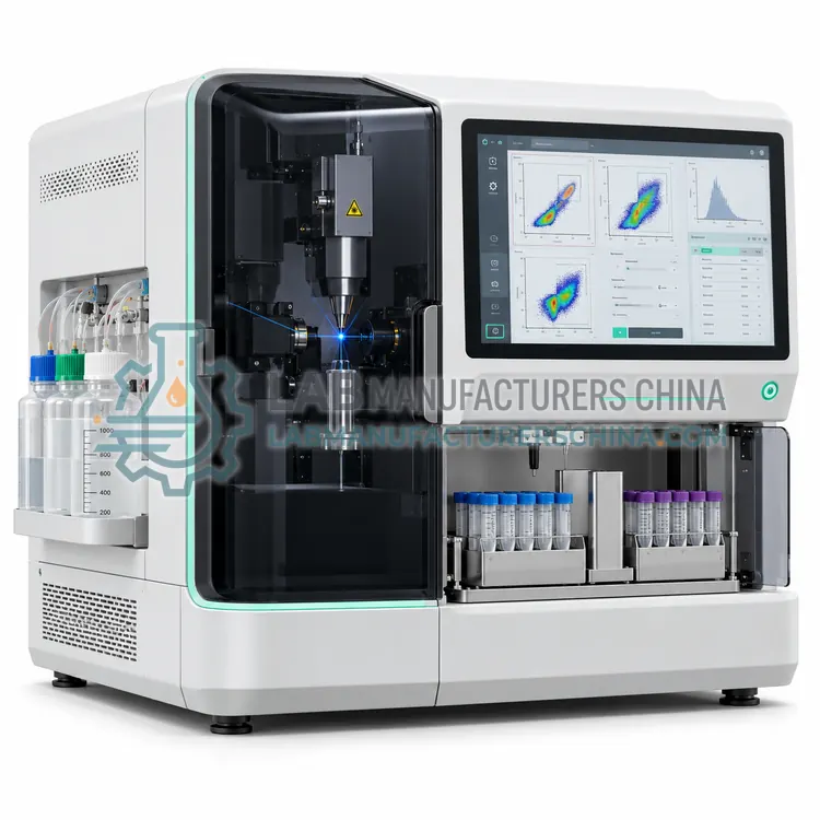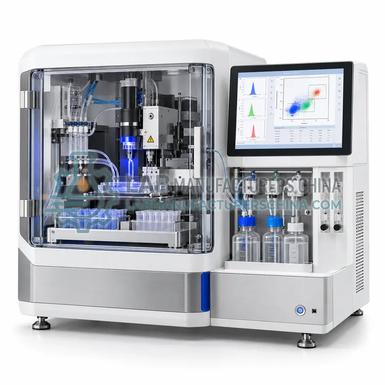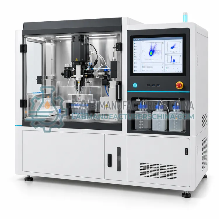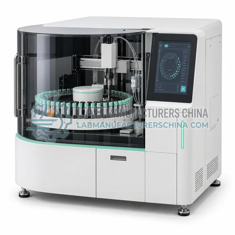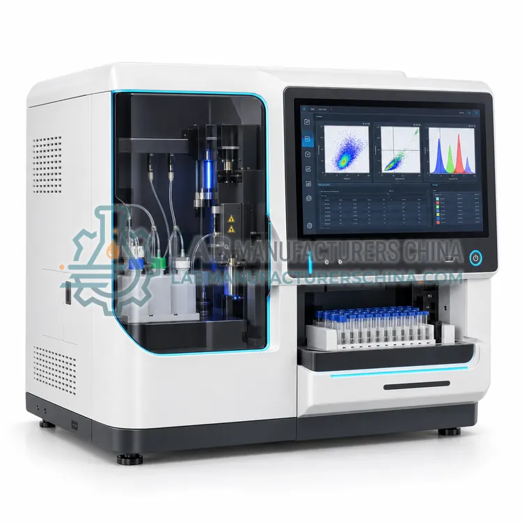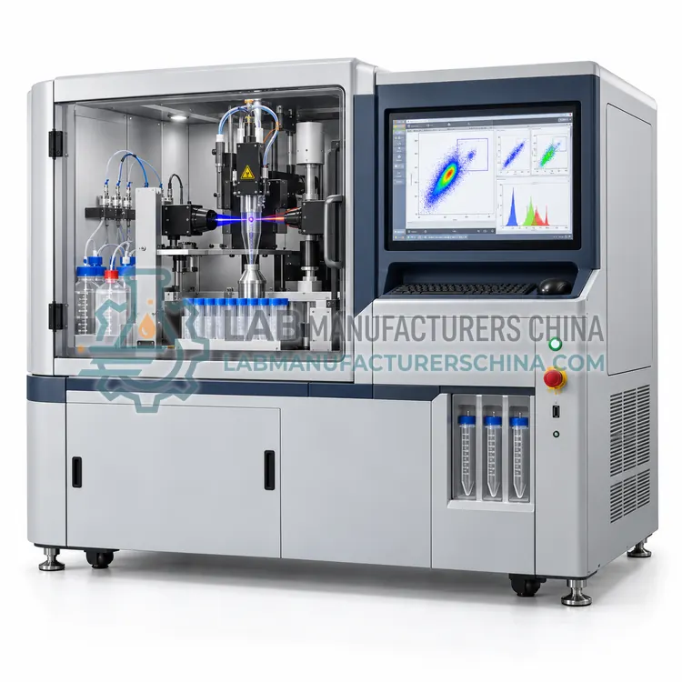
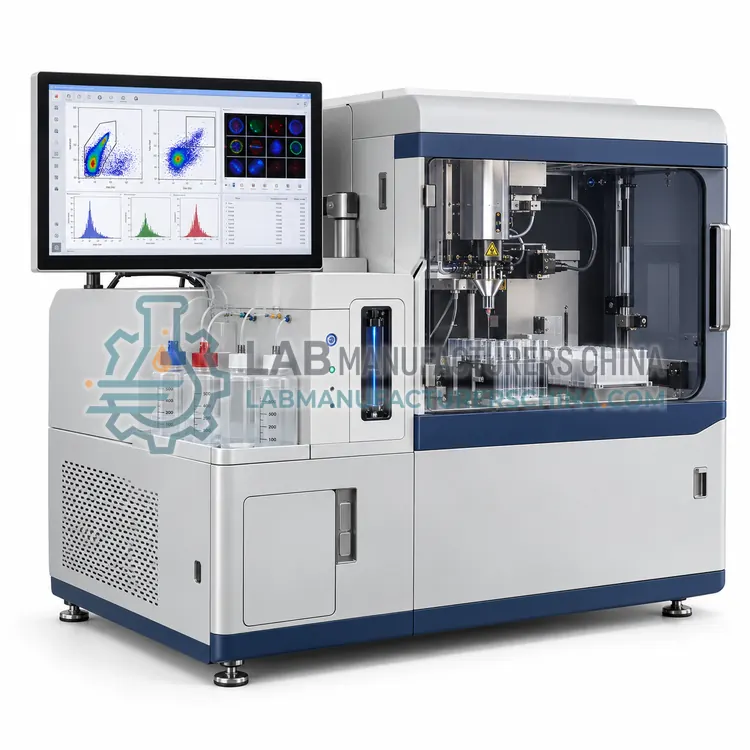
Image-Enabled Cell Sorter
成像型细胞分选仪
- SKU CSO-0005
- Cell Sorters
- CE (to be confirmed)
- IEC 61010-1 compliance (to be confirmed)
- IEC 60825-1 laser classification (to be confirmed)
Price on request — FOB / CIF quoted per order
Prices depend on configuration, quantity and destination port.
Key specifications
| Imaging modes | Brightfield plus up to 4 fluorescence |
|---|---|
| Image resolution | Approx. 0.5–1 µm per pixel |
| Laser configuration | 2–4 (405/488/561/640 nm) |
| Maximum sort rate | 1,000–15,000 events/s |
| Sort streams | 2-way (typical) |
| Image-derived features | Area, circularity, localization, texture |
Request a quote Download brochure · to be provided
By Lab Manufacturers China Technical TeamLast reviewed Standards: ISAC Cell Sorter Biosafety Standards, IEC 61010-1, IEC 60825-1
What is the Image-Enabled Cell Sorter?
An image-enabled cell sorter captures a high-speed image of each cell and sorts based on morphology, localization or cell-cell interaction in addition to fluorescence intensity. It is chosen when phenotypes cannot be resolved by intensity alone, such as nuclear translocation or doublet discrimination.
What is the Image-Enabled Cell Sorter?
An image-enabled cell sorter captures a high-speed image of each cell and sorts based on morphology, localization or cell-cell interaction in addition to fluorescence intensity. It is chosen when phenotypes cannot be resolved by intensity alone, such as nuclear translocation or doublet discrimination.
Overview
Image-enabled sorters combine flow cytometry with high-speed imaging, using time-delay integration cameras, optical time-stretch or frequency-encoded methods to acquire an image of every cell as it passes the interrogation point. Image-derived features such as cell area, nuclear-to-cytoplasmic ratio, protein localization, organelle count, or contact between cells are computed in real time and used as sorting parameters alongside scatter and fluorescence. Throughput is lower than conventional sorters, often 1,000 to 15,000 events per second, because of data volume and processing. Sorting may be droplet-based or via an integrated microfluidic actuator. These instruments enable spatial phenotypes to be linked to genotype through sorting followed by sequencing, and require specialized analysis software and trained users to build gating from image features.
How does it work?
Sorters analyze cells like an analyzer, then apply a decision in real time. In droplet sorters, a piezoelectric transducer vibrates the nozzle at 20 to 100 kHz so the stream breaks into uniform droplets just after the laser intercept. When a cell matching the sort gate is about to be enclosed, the electronics apply a charge to the stream, and the droplet separates carrying that charge. Charged droplets pass through high-voltage deflection plates and fall into tubes or multiwell plates, while uncharged droplets go to waste. A drop delay calibrated with fluorescent beads defines the time between detection and droplet break-off. Microfluidic chip sorters instead use valves, piezo actuators or optical forces inside a disposable cartridge, enclosing the sample away from aerosols.
What are the key features?
- High-speed imaging of every cell in brightfield and fluorescence channels
- Real-time image-derived features available as sort gating parameters
- Sort by morphology, protein localization or cell-cell interaction
- Doublet and debris discrimination improved by image-based gating
- Index sorting links image features to downstream sequencing data
- Dedicated software for image feature extraction and machine-learning classification
What are the technical specifications?
A typical image-enabled cell sorter configuration offers imaging modes of Brightfield plus up to 4 fluorescence, image resolution of Approx. 0.5–1 µm per pixel, laser configuration of 2–4 (405/488/561/640 nm), maximum sort rate of 1,000–15,000 events/s, sort streams of 2-way (typical) and image-derived features of Area, circularity, localization, texture. Values are indicative of the product class; the exact figures for each configuration are confirmed on the quotation and the calibration documents supplied with the instrument.
Which applications is it used for?
The Image-Enabled Cell Sorter is used in sorting by protein localization, nuclear translocation and signaling studies, cell-cell interaction and doublet analysis, morphology-based sorting for sequencing and phagocytosis and internalization assays. Across the wider Cell Sorters class, laboratories also deploy it for stem cell and progenitor enrichment, single-cell cloning of engineered cell lines and sorting for single-cell RNA sequencing.
Which standards and certifications apply?
Relevant standards and methods include ISAC Cell Sorter Biosafety Standards, IEC 61010-1, IEC 60825-1, EN 12469, ISO 14698-1, CLSI H62 and ISAC FCS 3.1. Certification candidates for this model are CE, IEC 61010-1 compliance and IEC 60825-1 laser classification; each is to be confirmed for the specific configuration before shipment, and certificates are supplied only where they have been issued.
Sorters need a clean, temperature-stable room, compressed air or nitrogen supply and often a dedicated operator. Perform daily setup with calibration beads to set drop delay, verify stream stability and check sort accuracy. Decontaminate fluidics with bleach or dedicated cleaning solutions before and after every sample and sterilize sheath paths for primary cell work. Sorting human or infectious material requires a biosafety assessment, with containment verified by an aerosol test such as potassium iodide or fluorescent bead methods. Nozzles, O-rings and sheath filters are consumables and should be replaced on a schedule. Laboratories should log sorts, purity checks and preventive maintenance to support reproducibility and audits.
How do I select the right Image-Enabled Cell Sorter?
Choose an image-enabled sorter only when the phenotype of interest depends on spatial or morphological information, since it runs slower and costs more than intensity-based sorters. In an RFQ, specify the imaging channels, optical resolution, sort rate, available image features and data storage and analysis requirements.
How does it compare with other Cell Sorters variants?
Choose a sorter on purity, yield, viability and containment requirements. Determine how many populations you need to collect at once, since two-way, four-way and six-way sorting require different deflection hardware. Match nozzle size, from 70 to 130 micrometers, to cell size and fragility; larger nozzles and lower pressure are gentler for primary cells. Check sort speed, typically 20,000 to 70,000 events per second, against the abort rate and recovery. For infectious, human primary or viral vector samples, specify an aerosol-managed or enclosed system and a class II biosafety cabinet. Consider single-cell deposition into 96 or 384-well plates, index sorting, number of lasers, and ease of use. Also confirm sterile fluidics cleaning and availability of service engineers.
Full specifications
| Imaging modes | Brightfield plus up to 4 fluorescence |
|---|---|
| Image resolution | Approx. 0.5–1 µm per pixel |
| Laser configuration | 2–4 (405/488/561/640 nm) |
| Maximum sort rate | 1,000–15,000 events/s |
| Sort streams | 2-way (typical) |
| Image-derived features | Area, circularity, localization, texture |
| Sort purity | Above 90% (typical) |
| Data storage per run | Tens of GB (image files) |
| Cell size range | Approx. 3–40 µm |
| Software | Image feature extraction and gating |
Indicative values for this product class. Final specifications are confirmed on the quotation and in the calibration documents supplied with the instrument.
Applications
- Sorting by protein localization
- Nuclear translocation and signaling studies
- Cell-cell interaction and doublet analysis
- Morphology-based sorting for sequencing
- Phagocytosis and internalization assays
Key features
- High-speed imaging of every cell in brightfield and fluorescence channels
- Real-time image-derived features available as sort gating parameters
- Sort by morphology, protein localization or cell-cell interaction
- Doublet and debris discrimination improved by image-based gating
- Index sorting links image features to downstream sequencing data
- Dedicated software for image feature extraction and machine-learning classification
Frequently asked questions about Image-Enabled Cell Sorter
It can sort on spatial information such as protein localization, nuclear translocation, organelle count or cells in contact, because it captures an image of each cell. Standard sorters rely only on integrated intensity and scatter signals.
Yes. Processing image data in real time limits throughput to roughly 1,000 to 15,000 events per second, well below conventional sorters. Rare populations may therefore need pre-enrichment or longer run times to collect enough cells.
Storing images for every cell creates large files, often tens of gigabytes per run, depending on channel count and event number. Plan for fast local storage, network backup and software that can compute features on the fly instead of saving all images.
It analyzes each cell by scatter and fluorescence, then a vibrating nozzle breaks the stream into droplets. Droplets containing cells that meet the sort gate are electrically charged and deflected by high-voltage plates into collection tubes or plates, leaving unwanted cells in waste.
Purity above 95 percent is common for well-separated populations sorted in purity mode, while yield and recovery are lower at high event rates. Actual results depend on gating, sample quality, sort mode, nozzle size and the abundance of target cells.
Sorting applies shear, pressure and electrostatic stress, so some loss of viability is possible. Using larger nozzles, lower sheath pressure, cooled collection, protein-supplemented collection media and lower event rates can preserve viability and downstream function for fragile primary cells.
Related models