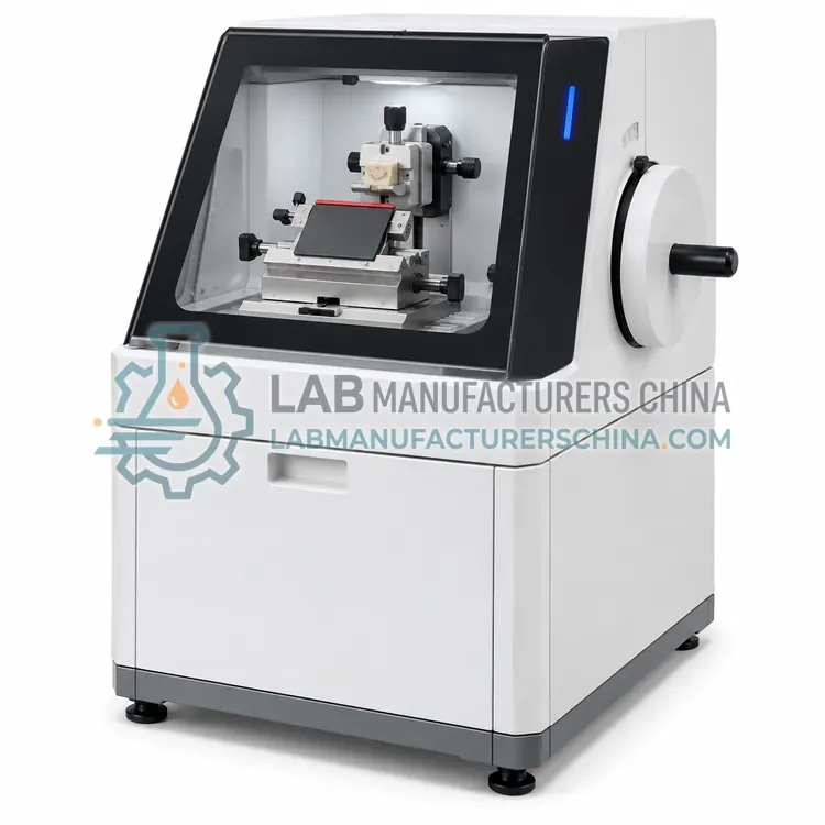
Rotary Microtome for Hard Tissue Sectioning
硬组织轮转式切片机
- SKU RMI-0005
- Rotary Microtomes
- CE (to be confirmed)
- IEC 61010-1 compliance (to be confirmed)
- ISO 13485 manufacturing (to be confirmed)
Price on request — FOB / CIF quoted per order
Prices depend on configuration, quantity and destination port.
Key specifications
| Section thickness range | 1–60 µm |
|---|---|
| Thickness increment | 1 µm |
| Maximum specimen size | about 60 × 50 mm |
| Specimen stroke | about 70 mm |
| Blade type | Tungsten carbide or heavy knife |
| Clamping force | Adjustable, higher than routine models |
Request a quote Download brochure · to be provided
By Lab Manufacturers China Technical TeamLast reviewed Standards: IEC 61010-1, IEC 61010-2-101, IEC 61326-1
What is the Rotary Microtome for Hard Tissue Sectioning?
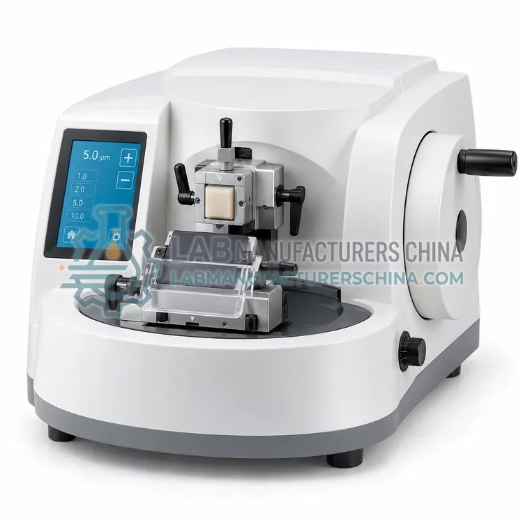
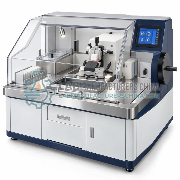
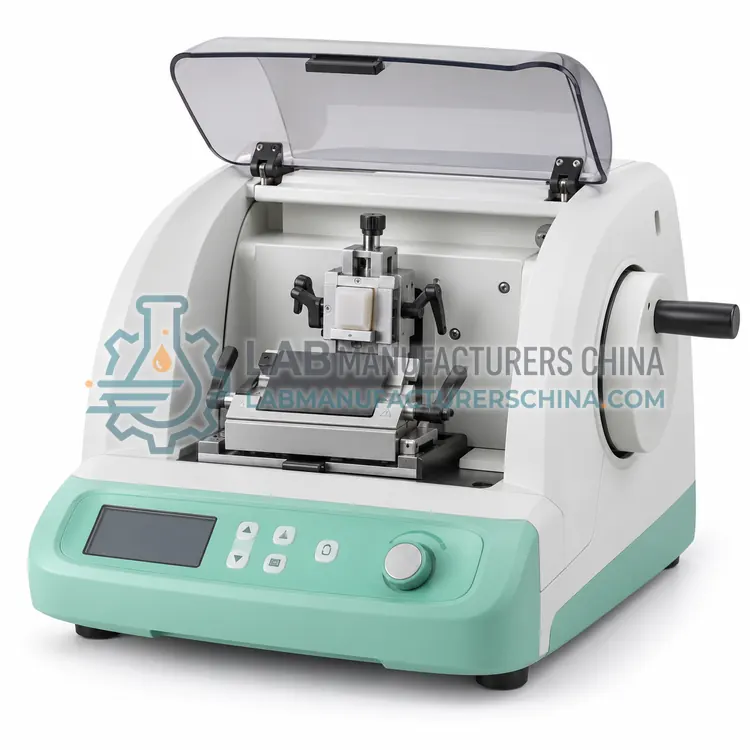
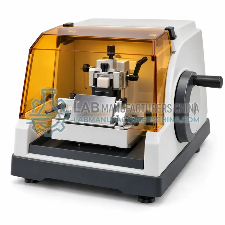
A hard tissue rotary microtome has a reinforced feed and blade holder for dense specimens such as undecalcified bone, teeth and implants. It is chosen when routine paraffin instruments cannot hold thickness or blade life.
What is the Rotary Microtome for Hard Tissue Sectioning?
A hard tissue rotary microtome has a reinforced feed and blade holder for dense specimens such as undecalcified bone, teeth and implants. It is chosen when routine paraffin instruments cannot hold thickness or blade life.
Overview
Hard tissue sectioning pushes a microtome beyond routine paraffin work. Bone, teeth, calcified tissue, biomaterials and metal implants resist cutting, so these instruments use stiffer feed mechanisms, larger blade or tungsten carbide knife holders, higher clamping force and sometimes a motorised feed to maintain thickness accuracy. Section thickness typically spans 1 to 60 µm for resin-embedded specimens, and some models cut thin ground sections from undecalcified bone at 5 to 10 µm with a tungsten carbide blade. Many laboratories combine resin embedding with a low-speed saw for initial sectioning, then finish on a hard tissue microtome. Cooling or moistening the block face and precise blade angle control reduce heat and chatter. Careful specimen fixation and infiltration are essential, because no instrument compensates for poor embedding.
How does it work?
A rotary microtome clamps a paraffin block in a cassette holder mounted on a feed mechanism and advances it toward a fixed blade as the operator turns a handwheel or a motor drives a cutting cycle. Each revolution advances the block by the set section thickness, usually adjustable from 1 to 60 µm in 1 µm steps, while a retraction mechanism pulls the block slightly back on the return stroke to protect the block face. Cut sections join into a continuous ribbon that is floated onto a warm water bath. Precision depends on rigid castings, low-backlash feed screws or stepper motors, a sharp disposable blade held at a fixed clearance angle and vibration-free installation. Blade angle, block orientation, ambient temperature and paraffin hardness also influence section quality, and coarse feed is used only for trimming.
What are the key features?
- Reinforced feed mechanism for dense specimens
- Tungsten carbide blade or heavy knife holder
- Higher clamping force and rigid block holder
- Section thickness about 1 to 60 µm for resin blocks
- Motorised feed option for thickness consistency
- Block face cooling or moistening accessory available
What are the technical specifications?
A typical rotary microtome for hard tissue sectioning configuration offers section thickness range of 1–60 µm, thickness increment of 1 µm, maximum specimen size of about 60 × 50 mm, specimen stroke of about 70 mm, blade type of Tungsten carbide or heavy knife and clamping force of Adjustable, higher than routine models. Values are indicative of the product class; the exact figures for each configuration are confirmed on the quotation and the calibration documents supplied with the instrument.
Which applications is it used for?
The Rotary Microtome for Hard Tissue Sectioning is used in undecalcified bone histology, dental and implant research, biomaterial and polymer implants and calcified tissue in veterinary studies. Across the wider Rotary Microtomes class, laboratories also deploy it for surgical pathology, research histology and immunohistochemistry preparation.
Which standards and certifications apply?
Relevant standards and methods include IEC 61010-1, IEC 61010-2-101, IEC 61326-1, ISO 15189 and ISO 13485. Certification candidates for this model are CE, IEC 61010-1 compliance and ISO 13485 manufacturing; each is to be confirmed for the specific configuration before shipment, and certificates are supplied only where they have been issued.
Install the microtome on a rigid bench away from vibration and drafts, in a room kept near 20 to 24 °C, because warm paraffin cuts poorly. Keep blades in a locked dispenser and dispose of used blades in a sharps container. Clean debris from the blade holder and block clamp after each session, lubricate guides as recommended, and check section thickness with a calibrated gauge or test block at defined intervals. Send instruments for professional service yearly. Record maintenance, calibration and training, and validate section quality through routine quality control blocks.
How do I select the right Rotary Microtome for Hard Tissue Sectioning?
Specify a hard tissue microtome when routine instruments produce cracked, compressed or under-thickness sections from resin blocks, bone or implants. Plan the whole workflow, because a low-speed saw and resin infiltration are usually needed as well. In the RFQ state maximum block size, blade or knife type, feed drive, cooling options and expected section thickness.
How does it compare with other Rotary Microtomes variants?
Choose rotary microtomes mainly by throughput and operator workload. Manual units are economical for low-volume laboratories that value tactile control. Semi-automatic models motorise the cutting stroke while leaving block advance to the operator, reducing fatigue at moderate volumes. Fully automatic instruments program cutting speed, thickness and section count for high-throughput or repetitive work. Motorised models with motorised specimen advance suit serial sectioning and hard tissue. When blocks are dense, such as undecalcified bone or calcified material, specify a version with reinforced feed and a tungsten carbide blade holder. In an RFQ state the thickness range, maximum block size, retraction, trimming function, blade system and whether a cooling stage is required.
Full specifications
| Section thickness range | 1–60 µm |
|---|---|
| Thickness increment | 1 µm |
| Maximum specimen size | about 60 × 50 mm |
| Specimen stroke | about 70 mm |
| Blade type | Tungsten carbide or heavy knife |
| Clamping force | Adjustable, higher than routine models |
| Feed drive | Manual or motorised |
| Cooling option | Block face cooling or moistening |
| Weight | about 40–55 kg |
Indicative values for this product class. Final specifications are confirmed on the quotation and in the calibration documents supplied with the instrument.
Applications
- Undecalcified bone histology
- Dental and implant research
- Biomaterial and polymer implants
- Calcified tissue in veterinary studies
Key features
- Reinforced feed mechanism for dense specimens
- Tungsten carbide blade or heavy knife holder
- Higher clamping force and rigid block holder
- Section thickness about 1 to 60 µm for resin blocks
- Motorised feed option for thickness consistency
- Block face cooling or moistening accessory available
Frequently asked questions about Rotary Microtome for Hard Tissue Sectioning
Yes, if the bone is resin-embedded, the instrument has a reinforced feed and a tungsten carbide blade is used. Sections of roughly 5 to 10 µm can be produced. Very dense or metal-containing specimens may require a low-speed saw for initial cutting.
Mineralised tissue and implants are abrasive and conduct heat poorly, so the cutting edge wears or softens rapidly. Tungsten carbide blades resist wear better than steel, and cutting speed and section thickness should be kept moderate to limit heat and edge damage.
For many hard tissue workflows it is. The saw produces the initial thin slice and the microtome refines thickness and surface quality. Some laboratories use only a sawing microtome, so the workflow should be planned before the instrument is purchased.
It cuts thin, uniform paraffin sections for light microscopy in histology. Sections are typically 1 to 60 µm thick and are used for routine H&E staining, special stains, immunohistochemistry and research, forming the basis of most pathology diagnosis.
Choose manual for low volumes and fine tactile control, semi-automatic when cutting stroke fatigue is a concern, and fully automatic for high throughput or repetitive serial sectioning. Automatic models also give more reproducible speed and thickness, which supports consistent section quality.
Curling and wrinkling usually come from a blunt or dirty blade, a wrong clearance angle, paraffin that is too cold or too soft, or too fast a cutting stroke. Check blade angle, block temperature and cutting speed, and float sections on a water bath at about 40 to 45 °C.
Related models