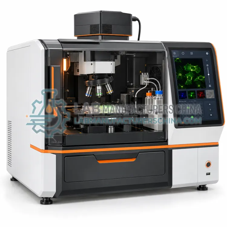
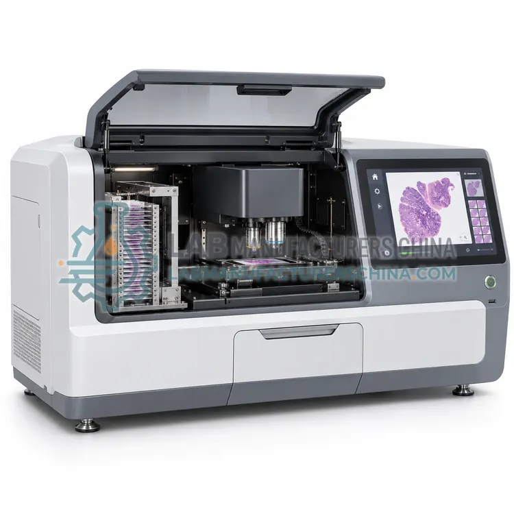
Whole-Slide Scanning Imaging System
全切片扫描成像系统
- SKU ISY-0004
- Imaging Systems
- CE (to be confirmed)
- ISO 13485 manufacturing (to be confirmed)
- IVDR (EU) 2017/746 (to be confirmed)
Price on request — FOB / CIF quoted per order
Prices depend on configuration, quantity and destination port.
Key specifications
| Slide capacity | 1-400 slides per load |
|---|---|
| Magnification | 20x and 40x objectives |
| Resolution | 0.25 µm/px at 20x, 0.12 µm/px at 40x |
| Imaging modes | Brightfield standard, 3-4 fluorescence channels optional |
| Scan time | 1-5 min per slide |
| Focus | Focus map with z-stack option |
Request a quote Download brochure · to be provided
By Lab Manufacturers China Technical TeamLast reviewed Standards: IEC 61010-1, IEC 61326-1, ISO 9022-1
What is the Whole-Slide Scanning Imaging System?
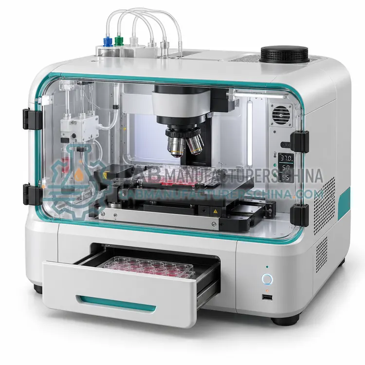
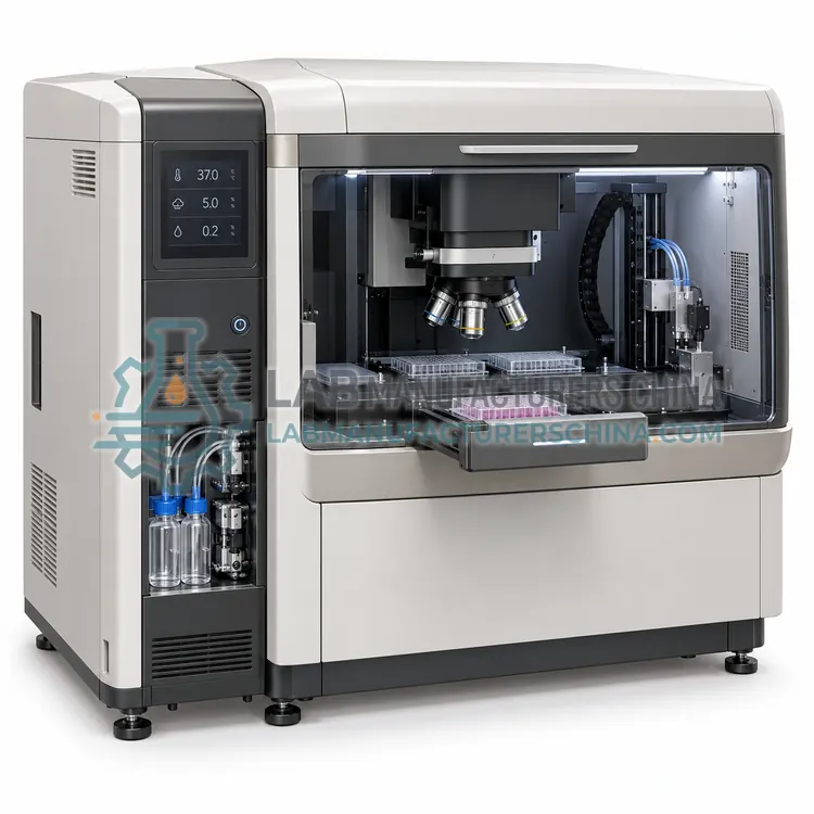
A slide scanner that digitizes entire stained tissue sections at 20x or 40x magnification into stitched, high-resolution image files. It supports digital pathology review, quantitative image analysis and remote consultation between clinical sites.
What is the Whole-Slide Scanning Imaging System?
A slide scanner that digitizes entire stained tissue sections at 20x or 40x magnification into stitched, high-resolution image files. It supports digital pathology review, quantitative image analysis and remote consultation between clinical sites.
Overview
The whole-slide scanner loads standard 25 by 75 mm slides into a magazine of 1 to 400 slides, then tiles the entire section using a motorized stage and line-scan or area-scan camera. Brightfield scanning with a white LED is standard, and fluorescence channels are available on research models. Resolution is defined by numerical aperture and pixel spacing, typically 0.25 µm per pixel at 20x and 0.12 µm per pixel at 40x, and focus maps followed by z-stacking keep uneven sections sharp. Scan time ranges from one to five minutes per slide depending on magnification and tissue area, and images are stored in vendor or open whole-slide formats with compressed pyramids for fast viewing. Barcode recognition, case lists and integration with laboratory information systems support routine pathology workflows.
How does it work?
Most life science imaging systems share the same optical path: an excitation source such as an LED, laser or arc lamp sends light through filters or a monochromator to the sample, and an objective collects the emitted light, which a cooled CCD or sCMOS camera converts into digital counts. Fluorescence depends on fluorophores that absorb at one wavelength and emit at a longer one, so filter cubes or tunable filters separate excitation from emission. Brightfield imaging relies on transmitted light and contrast techniques such as phase contrast or differential interference contrast. Motorized stages, autofocus routines and multi-well plate carriers let the system visit many positions in a programmed order, and software assembles the images, applies flat-field and dark corrections, and extracts measurements such as intensity, area, count and morphology.
What are the key features?
- Slide magazine for 1 to 400 standard slides.
- Brightfield scanning with optional fluorescence channels.
- Resolution from 0.25 to 0.12 µm per pixel.
- Focus mapping and z-stacking for uneven sections.
- Scan time of one to five minutes per slide.
- Barcode reading and laboratory information system integration.
What are the technical specifications?
A typical whole-slide scanning imaging system configuration offers slide capacity of 1-400 slides per load, magnification of 20x and 40x objectives, resolution of 0.25 µm/px at 20x, 0.12 µm/px at 40x, imaging modes of Brightfield standard, 3-4 fluorescence channels optional, scan time of 1-5 min per slide and focus of Focus map with z-stack option. Values are indicative of the product class; the exact figures for each configuration are confirmed on the quotation and the calibration documents supplied with the instrument.
Which applications is it used for?
The Whole-Slide Scanning Imaging System is used in digital pathology slide archiving, remote pathology consultation, quantitative tissue image analysis and research histology and immunohistochemistry scanning. Across the wider Imaging Systems class, laboratories also deploy it for fluorescence and immunofluorescence microscopy, live-cell dynamics and time-lapse recording and high-content screening and phenotypic assays.
Which standards and certifications apply?
Relevant standards and methods include IEC 61010-1, IEC 61326-1, ISO 9022-1, ISO 13485 manufacturing and IVDR (EU) 2017/746. Certification candidates for this model are CE, ISO 13485 manufacturing and IVDR (EU) 2017/746; each is to be confirmed for the specific configuration before shipment, and certificates are supplied only where they have been issued.
Imaging performance depends on the room as much as on the instrument. Use a vibration-isolated bench away from centrifuges and pumps, keep temperature stable, and limit stray light for long exposures or bioluminescence work. Provide stable power, ideally through a line conditioner, and a network path for image data, which can exceed tens of gigabytes per session. Calibrate the stage, objective magnification and fluorescence channels with reference slides at installation and at defined intervals, and verify camera linearity and dark current. Clean objectives with lens tissue and approved solvent only. Manage data with a documented naming and backup policy, since files are large and often subject to retention requirements.
How do I select the right Whole-Slide Scanning Imaging System?
Select the slide scanner when whole sections must be reviewed on screen, archived digitally or measured quantitatively. Compare slide capacity and scan time against daily volume, and check magnification, resolution and whether fluorescence channels are needed. Confirm the file format, viewer licensing, image-analysis compatibility and laboratory information system integration before ordering.
How does it compare with other Imaging Systems variants?
Match the system to the question: fixed-cell morphology and staining favour an inverted fluorescence microscope platform, while dynamic processes need an incubator-enclosed live-cell system. Throughput decides between manual microscopes, plate-capable high-content readers and slide scanners, which differ mainly in automation, plate handling and analysis software. Detection sensitivity is the next filter, since weak luminescence needs deep-cooled cameras and strong fluorophores tolerate less cooling. Check objective range and numerical aperture, excitation channels, camera quantum efficiency and pixel size, stage resolution, autofocus reliability and the image format and analysis tools. Confirm that the software exports raw data and supports the statistics package your team uses.
Full specifications
| Slide capacity | 1-400 slides per load |
|---|---|
| Magnification | 20x and 40x objectives |
| Resolution | 0.25 µm/px at 20x, 0.12 µm/px at 40x |
| Imaging modes | Brightfield standard, 3-4 fluorescence channels optional |
| Scan time | 1-5 min per slide |
| Focus | Focus map with z-stack option |
| Illumination | White LED, stable output |
| Slide format | 25 mm × 75 mm, 1 mm thick |
| File formats | Vendor pyramid format and open whole-slide format |
| Data output | Network share with case list and barcode |
Indicative values for this product class. Final specifications are confirmed on the quotation and in the calibration documents supplied with the instrument.
Applications
- Digital pathology slide archiving
- Remote pathology consultation
- Quantitative tissue image analysis
- Research histology and immunohistochemistry scanning
Key features
- Slide magazine for 1 to 400 standard slides.
- Brightfield scanning with optional fluorescence channels.
- Resolution from 0.25 to 0.12 µm per pixel.
- Focus mapping and z-stacking for uneven sections.
- Scan time of one to five minutes per slide.
- Barcode reading and laboratory information system integration.
Frequently asked questions about Whole-Slide Scanning Imaging System
Choose 40x when cytological detail such as nuclear chromatin matters, and 20x when throughput is more important. Many laboratories scan at 20x for routine work and rescan selected slides at 40x, which saves storage and time.
A 20x brightfield slide typically occupies 0.5 to 2 GB. Scanning a few hundred slides a week quickly reaches several terabytes, so plan a network archive with backup and a retention policy before installation.
In many jurisdictions yes, provided the scanner is validated for the intended use, the display meets requirements and quality controls cover focus and colour. Check local regulatory expectations and keep the validation records with the instrument.
A microscope shows an image to the eye, while an imaging system adds a camera, motorised stage, controlled illumination and software that capture, store and quantify images. That combination enables repeated measurements, multi-well automation and statistical analysis across many samples.
Cooled sCMOS or back-illuminated CCD cameras with quantum efficiency above 80 percent and low read noise are preferred. Deep cooling reduces dark current during long exposures, which matters for weak signals such as bioluminescence or single-molecule fluorescence.
A single 96-well plate recorded in three channels can reach tens of gigabytes, and time-lapse or slide-scanning runs are larger. Plan network transfer speed, working storage and a backup or archival policy before the system is installed.
Related models