Microfluidic Chip Fragment Analyzer
微流控芯片片段分析仪
- SKU FRA-0003
- Fragment Analyzers
- CE (to be confirmed)
- ISO 9001 manufacturing (to be confirmed)
- IEC 61010-1 safety (to be confirmed)
Price on request — FOB / CIF quoted per order
Prices depend on configuration, quantity and destination port.
Key specifications
| Lanes per chip | 10–16 |
|---|---|
| Analysis time | approx. 25–40 min |
| Sample volume | 1–5 µL per lane |
| Sizing range | 25 bp–12 kb (kit dependent) |
| Sizing accuracy | within ±10% (typical) |
| Sensitivity | approx. 0.1–50 ng/µL (kit dependent) |
Request a quote Download brochure · to be provided
By Lab Manufacturers China Technical TeamLast reviewed Standards: ISO 20395, ISO 22174, CLSI MM09
What is the Microfluidic Chip Fragment Analyzer?
A microfluidic chip analyzer sizes and quantifies nucleic acids on a disposable chip with 10 to 16 lanes, using microlitre volumes and giving results in about 30 minutes. It suits laboratories that need fast, low-volume quality control of libraries, RNA and PCR products.
What is the Microfluidic Chip Fragment Analyzer?
A microfluidic chip analyzer sizes and quantifies nucleic acids on a disposable chip with 10 to 16 lanes, using microlitre volumes and giving results in about 30 minutes. It suits laboratories that need fast, low-volume quality control of libraries, RNA and PCR products.
Overview
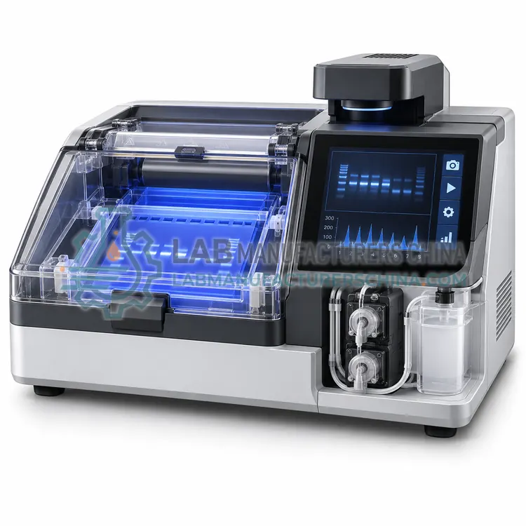
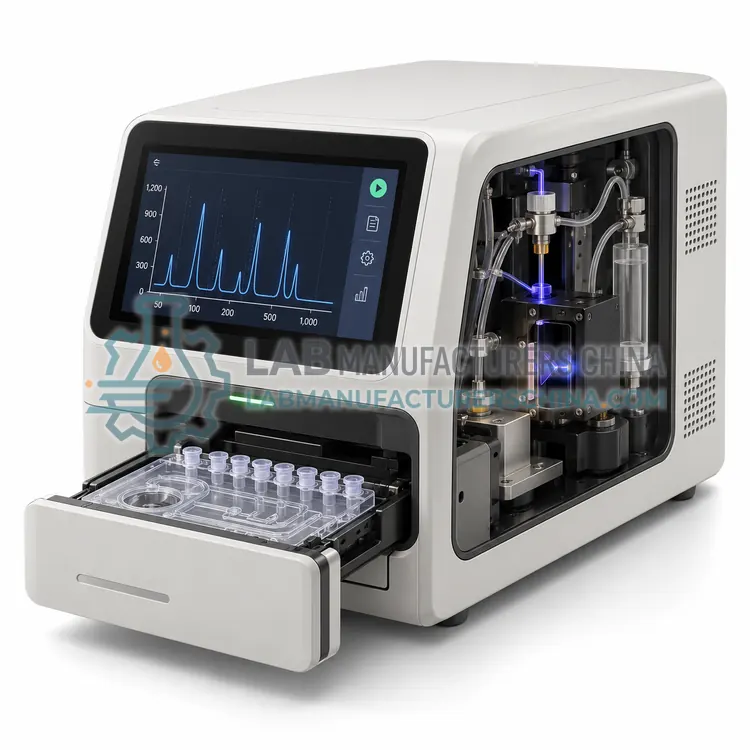
The chip contains etched channels, reservoirs and electrodes; samples, ladder and gel-dye mix are pipetted into wells and the chip is placed on the instrument, where electrodes drive electrokinetic injection and separation. Fluorescence detection records each lane, and software reports fragment size, concentration, molarity and RNA integrity. A 10- to 16-lane chip is normally completed in 25–40 minutes, which is faster than plate-based systems for small numbers of samples. The format uses 1–5 µL of sample, conserving low-input libraries. Resolution is suitable for detecting adapter dimers, primer peaks and degraded RNA, but sizing above about 12 kb and below 25 bp is limited. Chips are single-use, and consumable cost per sample is usually higher than for capillary plates.
How does it work?
Fragment analysis separates charged nucleic acid fragments in a sieving matrix and detects them by fluorescence. In capillary systems, samples are injected electrokinetically into a polymer-filled capillary, where smaller fragments migrate faster and reach the detector earlier. A laser excites intercalating dye bound to the DNA or RNA, and the software converts migration times into fragment sizes using an internal ladder and into concentrations using a quantitative marker. Microfluidic chips work on the same principle with shorter separation channels and smaller volumes. Automated agarose gel instruments draw samples through a gel by voltage and image the bands after staining. Because the sizing algorithm depends on the ladder and the polymer lot, run quality flags such as marker height and baseline noise should be reviewed before results are accepted.
What are the key features?
- Disposable chip with 10 to 16 sample lanes
- Results for a full chip in about 25–40 minutes
- Sample consumption of only 1–5 µL per lane
- Detects adapter dimers, primer peaks and degraded RNA
- Pre-mixed gel-dye kits reduce preparation steps
- Gel-like image and electropherogram views in one report
What are the technical specifications?
A typical microfluidic chip fragment analyzer configuration offers lanes per chip of 10–16, analysis time of approx. 25–40 min, sample volume of 1–5 µL per lane, sizing range of 25 bp–12 kb (kit dependent), sizing accuracy of within ±10% (typical) and sensitivity of approx. 0.1–50 ng/µL (kit dependent). Values are indicative of the product class; the exact figures for each configuration are confirmed on the quotation and the calibration documents supplied with the instrument.
Which applications is it used for?
The Microfluidic Chip Fragment Analyzer is used in quick library checks before sequencing, RNA integrity assessment of small sample sets, PCR product and primer dimer detection and low-input sample quality control. Across the wider Fragment Analyzers class, laboratories also deploy it for next-generation sequencing library quality control, RNA integrity assessment before expression studies and PCR amplicon and restriction digest checks.
Which standards and certifications apply?
Relevant standards and methods include ISO 20395, ISO 22174, CLSI MM09, IEC 61010-1 and ISO 15189. Certification candidates for this model are CE, ISO 9001 manufacturing and IEC 61010-1 safety; each is to be confirmed for the specific configuration before shipment, and certificates are supplied only where they have been issued.
Install the analyzer on a level bench away from vibration and direct sunlight, because optical alignment and polymer flow are sensitive to movement. Store polymer, ladders and dye kits within their stated temperature range and note lot numbers, since a ladder or polymer change can shift sizing by several percent. Run a ladder and a known control with each batch, and review quality flags before releasing data. Replace polymer and clean electrodes as instructed to prevent carry-over between samples. Keep the instrument in a room with stable temperature to limit baseline drift, and include the analyzer in the laboratory's pipette and instrument calibration schedule. Verify performance after any service or software upgrade.
How do I select the right Microfluidic Chip Fragment Analyzer?
Select a microfluidic chip analyzer when sample numbers are small to moderate, sample volume is limited and results are needed within an hour. Specify lane count, sizing range, kit sensitivity, consumable cost per chip and whether the software reports RNA integrity and library region values.
How does it compare with other Fragment Analyzers variants?
Choose by the sample type, resolution and throughput you need. Capillary systems give the best sizing accuracy and reproducibility, which matters for sequencing libraries and RNA integrity scoring. Microfluidic chip systems run more samples in less time with microlitre volumes but give approximate sizing. Automated gel systems handle many samples at low consumable cost with lower resolution. Check the sizing range against your fragments: amplicons of 100–1,000 bp behave differently from long-read libraries above 20 kb, and high-sensitivity kits are needed below 1 ng/µL. Confirm sample volume, consumable cost, run time, and whether the software exports results to a laboratory information system. Where quantification must be exact, pair the analyzer with a fluorometric or digital PCR method.
Full specifications
| Lanes per chip | 10–16 |
|---|---|
| Analysis time | approx. 25–40 min |
| Sample volume | 1–5 µL per lane |
| Sizing range | 25 bp–12 kb (kit dependent) |
| Sizing accuracy | within ±10% (typical) |
| Sensitivity | approx. 0.1–50 ng/µL (kit dependent) |
| Detection | Laser-induced fluorescence |
| RNA assay range | approx. 25–6,000 nt |
| Chip format | Single-use microfluidic chip with prefilled gel-dye wells |
| Dimensions (W x D x H) | approx. 260 x 360 x 210 mm |
Indicative values for this product class. Final specifications are confirmed on the quotation and in the calibration documents supplied with the instrument.
Applications
- Quick library checks before sequencing
- RNA integrity assessment of small sample sets
- PCR product and primer dimer detection
- Low-input sample quality control
Key features
- Disposable chip with 10 to 16 sample lanes
- Results for a full chip in about 25–40 minutes
- Sample consumption of only 1–5 µL per lane
- Detects adapter dimers, primer peaks and degraded RNA
- Pre-mixed gel-dye kits reduce preparation steps
- Gel-like image and electropherogram views in one report
Frequently asked questions about Microfluidic Chip Fragment Analyzer
Chips are faster for small batches and use less sample, but cost more per sample and offer a narrower sizing range with slightly lower accuracy. Capillary plates suit larger batches and long-read libraries.
Yes. Adapter dimers appear as a peak near 120–180 bp, well below the library insert size, and the software can quantify the percentage of total signal in that region so poor libraries are identified before sequencing.
No. Channels are loaded with gel and dye and cannot be cleaned reliably, so chips are single-use consumables. Electrodes on the instrument are cleaned between runs following the supplier procedure.
It reports the size in base pairs or nucleotides and the concentration of each fragment in a sample, along with a size distribution. For RNA it also computes an integrity score. Protein assays report molecular weight and relative purity.
Capillary systems give better sizing accuracy, quantitative concentration and digital results that can be exported and audited. Gels are cheaper per sample and show many samples at once but need manual interpretation and are less reproducible for sizing below 5% differences.
Run the RNA on a capillary or chip system that reports an integrity score computed from the whole electropherogram, including ribosomal peaks and degradation products. Many sequencing and expression workflows require a minimum score and reject samples below it.