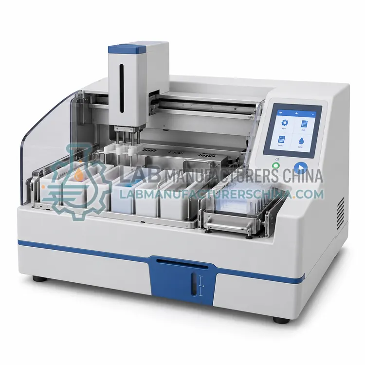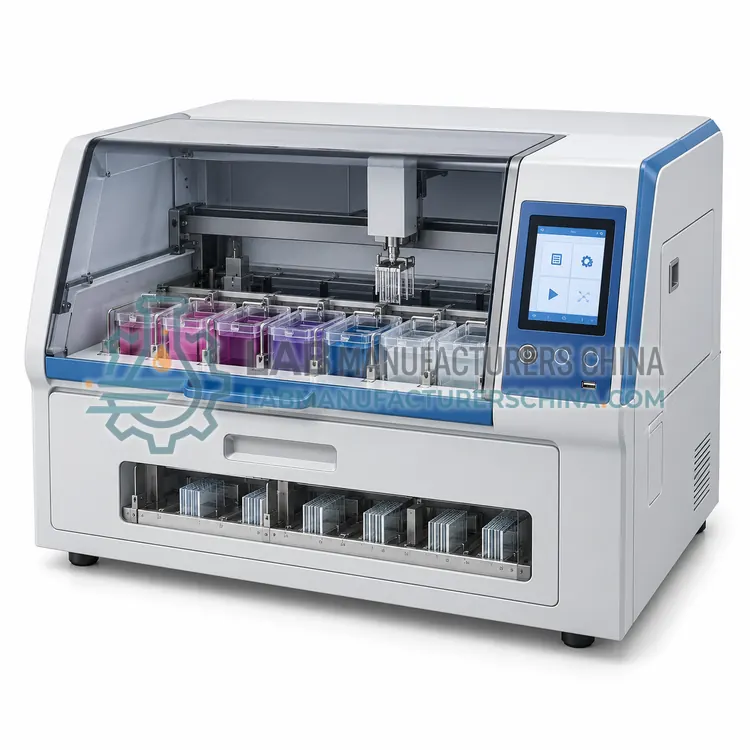
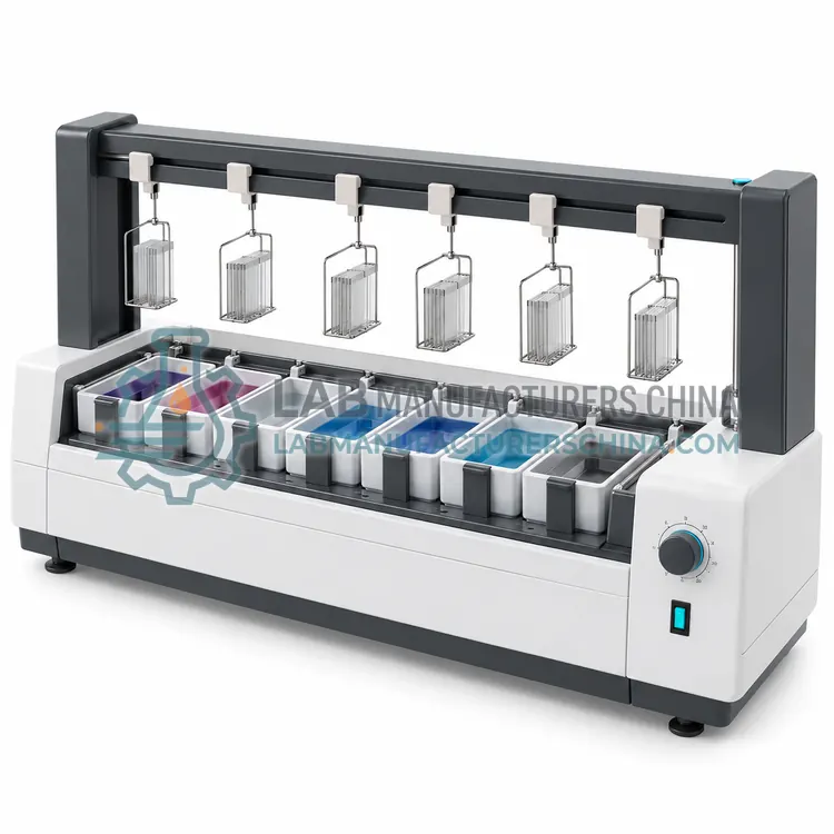
Manual Cytology Staining Station
手工细胞学染色工作站
- SKU CST-0001
- Cytology Stainers
- CE (to be confirmed)
- IEC 61010-1 compliance (to be confirmed)
- ISO 13485 manufacturing (to be confirmed)
Price on request — FOB / CIF quoted per order
Prices depend on configuration, quantity and destination port.
Key specifications
| Staining bath count | 10–20 baths |
|---|---|
| Bath volume | about 200–300 mL |
| Slide capacity per rack | 20–30 slides |
| Rinse station | Running water or buffer |
| Exhaust airflow | about 0.3–0.5 m/s face velocity |
| Bench material | Solvent-resistant phenolic resin |
Request a quote Download brochure · to be provided
By Lab Manufacturers China Technical TeamLast reviewed Standards: IEC 61010-1, IEC 61010-2-101, IEC 61326-1
What is the Manual Cytology Staining Station?
A manual cytology staining station groups staining baths, rinse stations and a fume enclosure for operator-controlled staining. It suits low-volume cytology work, teaching and research protocols that need careful visual judgement of colour development.
What is the Manual Cytology Staining Station?
A manual cytology staining station groups staining baths, rinse stations and a fume enclosure for operator-controlled staining. It suits low-volume cytology work, teaching and research protocols that need careful visual judgement of colour development.
Overview
The manual station is the reference method for cytology staining and remains useful wherever volumes are low or protocols vary. It typically comprises a rack of staining baths for hematoxylin, orange G, eosin-azure and alcohols, a rinse area, and local exhaust ventilation for any solvent-containing steps. Technicians move slide racks by hand and judge colour development visually, adjusting dwell times to compensate for aging reagents. This flexibility is valuable for research methods and for unusual sample types that do not behave like cervical smears. The disadvantages are variability between operators, exposure to dyes and alcohols, and the labour required for large batches. Timed baths, a written schedule and control slides help reduce variation, and a solvent-resistant bench with spill containment is essential.
How does it work?
Cytology staining differentiates cellular structures by exploiting their affinity for dyes. In the Papanicolaou method, hematoxylin stains nuclei blue, while orange G and eosin-azure counterstains give keratinised and metabolically active cells their characteristic orange, pink and blue-green shades. Giemsa-type stains give a panoptic result that shows nuclear chromatin, cytoplasm and microorganisms, and is common for air-dried samples. A stainer moves slide racks or individual slides through alcohol, dye, rinse and clearing stations for controlled times, often with agitation, and may dry them before coverslipping. Alcohol concentrations, dye pH, dwell time and rinse efficiency determine the final balance of colours, so reagent quality and stable timing are the critical variables.
What are the key features?
- Rack of staining baths for dye and alcohol steps
- Rinse area for running water or buffer
- Local exhaust ventilation for solvent steps
- Operator control of dwell time and colour
- Low cost and flexible for unusual protocols
- Suited to teaching and method development
What are the technical specifications?
A typical manual cytology staining station configuration offers staining bath count of 10–20 baths, bath volume of about 200–300 mL, slide capacity per rack of 20–30 slides, rinse station of Running water or buffer, exhaust airflow of about 0.3–0.5 m/s face velocity and bench material of Solvent-resistant phenolic resin. Values are indicative of the product class; the exact figures for each configuration are confirmed on the quotation and the calibration documents supplied with the instrument.
Which applications is it used for?
The Manual Cytology Staining Station is used in low-volume cytology, teaching and training, research cytology protocols and unusual sample types. Across the wider Cytology Stainers class, laboratories also deploy it for cervical cytology screening, papanicolaou staining and giemsa staining of air-dried samples.
Which standards and certifications apply?
Relevant standards and methods include IEC 61010-1, IEC 61010-2-101, IEC 61326-1 and ISO 15189. Certification candidates for this model are CE, IEC 61010-1 compliance and ISO 13485 manufacturing; each is to be confirmed for the specific configuration before shipment, and certificates are supplied only where they have been issued.
Install the stainer near water and drainage if rinsing requires running water, and keep it away from direct light that fades dyes. Prepare fresh hematoxylin and counterstains on a defined schedule based on slide counts, and monitor with control slides, because colour drift is usually chemical rather than mechanical. Clean baths, nozzles and tubing to prevent precipitate and microbial growth, and check that filtering steps remove dye particles. Record reagent lots, changes and quality control results. Train staff in safe handling of dyes and alcohols, and ensure adequate ventilation for any solvent-based clearing step.
How do I select the right Manual Cytology Staining Station?
Select a manual station for low volumes, teaching or method development where you must see and control colour development. Confirm adequate ventilation and a solvent-resistant bench, and plan for timed baths and control slides to limit variation between operators.
How does it compare with other Cytology Stainers variants?
Choose a cytology stainer by slide volume, stain menu and workflow. Manual staining stations suit low volumes and teaching, and give full control when colours need adjustment. Automated Papanicolaou stainers are the standard for cervical screening, where hundreds of slides per day must look identical; Giemsa stainers suit haematology and air-dried cytology. Linear stainers offer moderate throughput with simple mechanics, while benchtop units fit small laboratories. Compare slides per run, station count and volume, programmability, agitation, wash efficiency, drying and coverslipping integration. Check carryover prevention, reagent consumption and whether the instrument accepts third-party reagents or requires proprietary kits.
Full specifications
| Staining bath count | 10–20 baths |
|---|---|
| Bath volume | about 200–300 mL |
| Slide capacity per rack | 20–30 slides |
| Rinse station | Running water or buffer |
| Exhaust airflow | about 0.3–0.5 m/s face velocity |
| Bench material | Solvent-resistant phenolic resin |
| Timing aid | Interval timer or timed baths |
| Power supply | 220 V, 50 Hz |
Indicative values for this product class. Final specifications are confirmed on the quotation and in the calibration documents supplied with the instrument.
Applications
- Low-volume cytology
- Teaching and training
- Research cytology protocols
- Unusual sample types
Key features
- Rack of staining baths for dye and alcohol steps
- Rinse area for running water or buffer
- Local exhaust ventilation for solvent steps
- Operator control of dwell time and colour
- Low cost and flexible for unusual protocols
- Suited to teaching and method development
Frequently asked questions about Manual Cytology Staining Station
Yes, for low volumes, teaching and non-standard protocols, provided timing and reagent control are documented. For screening programmes with high daily volumes, automated staining gives more consistent colour and lower operator exposure.
Usually because reagents have aged, alcohol concentrations have shifted through evaporation, or rinse water quality has changed. Check pH, dye filtration and alcohol strength, and compare with a control slide stained at the same time.
Local exhaust ventilation for dye and alcohol steps, ideally a fume hood or down-draft bench with face velocity around 0.3 to 0.5 m/s. General room ventilation alone is usually insufficient for solvent-containing clearing steps.
It applies differential stains such as Papanicolaou or Giemsa to cell preparations, giving reproducible colour that shows nuclear and cytoplasmic detail. It replaces manual dipping so large numbers of slides stain consistently in screening and diagnostic laboratories.
Papanicolaou staining uses hematoxylin with orange G and eosin-azure counterstains and is designed for alcohol-fixed smears, giving clear nuclear detail and keratin colours. Giemsa staining is a panoptic stain suited to air-dried samples and is common in haematology.
Control reagent quality and timing. Prepare fresh dyes on a schedule based on slide counts, check pH and alcohol concentration, filter to remove precipitate, and run control slides each batch. Most colour drift is caused by exhausted reagents rather than the instrument.
Related models