Vibrating Microtome Tissue Slicer
振动切片机
- SKU CEP-0005
- Cell Processing
- CE (to be confirmed)
- ISO 9001 manufacturing (to be confirmed)
- IEC 61010-1 safety (to be confirmed)
Price on request — FOB / CIF quoted per order
Prices depend on configuration, quantity and destination port.
Key specifications
| Section thickness | 100–1,000 µm |
|---|---|
| Blade oscillation | 0.5–1.5 mm per stroke, up to 100 Hz |
| Advance speed | 0.01–2 mm/s |
| Cutting chamber volume | approx. 100–200 mL |
| Temperature control | 2–45 °C with circulating bath |
| Maximum specimen size | approx. 40 x 40 mm |
Request a quote Download brochure · to be provided
By Lab Manufacturers China Technical TeamLast reviewed Standards: IEC 61010-1, IEC 61010-2-010, IEC 61010-2-020
What is the Vibrating Microtome Tissue Slicer?
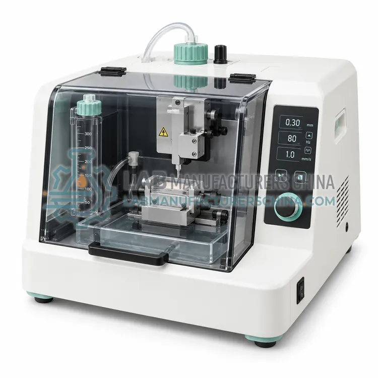
A vibrating microtome cuts living tissue into thin slices with a rapidly oscillating blade that minimises compression, preserving cell viability and local circuitry for electrophysiology, explant culture and live tissue imaging experiments.
What is the Vibrating Microtome Tissue Slicer?
A vibrating microtome cuts living tissue into thin slices with a rapidly oscillating blade that minimises compression, preserving cell viability and local circuitry for electrophysiology, explant culture and live tissue imaging experiments.
Overview
The blade moves laterally at high frequency, typically 0.5–1.5 mm per stroke, while the tissue is advanced slowly and submerged in cold, oxygenated buffer. Low advance speed and lateral oscillation mean that cutting forces are small, so cells near the slice surface remain viable and the cytoarchitecture is preserved. Section thickness of 100–500 µm covers most neural slice electrophysiology and explant work, and thicker sections of up to 1 mm are used for some preparations. Chambers are filled with carbogen-buffered artificial cerebrospinal fluid or culture medium, and temperature can be held near 4 °C for neural tissue or 37 °C for other applications. Magnetic or adhesive stage plates hold tissue securely without embedding, and blade advance rates are adjustable for soft or fibrous material.
How does it work?
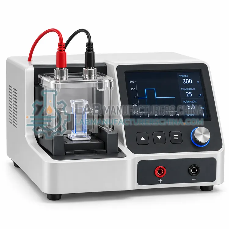
Each processing step addresses a different physical or biological barrier. Electroporation applies a brief electric pulse that transiently increases membrane permeability so plasmid DNA, RNA or protein can enter the cytoplasm or nucleus; efficiency depends on field strength, pulse shape and buffer conductivity. Thawing instruments warm frozen vials under controlled heat transfer so that cells pass quickly through the temperature range where ice recrystallisation damages membranes. Tissue dissociation combines enzymatic digestion with controlled mechanical shear, using a heater-shaker, a gentleMACS-style rotor or a closed automated chamber, to release single cells while limiting DNA release that causes clumping. Media exchange systems centrifuge, resuspend and wash cells in a closed circuit for concentration and formulation. Vibrating microtomes cut living tissue into slices with minimal damage, preserving neural circuitry for electrophysiology and explant culture.
What are the key features?
- Rapid lateral blade oscillation at 0.5–1.5 mm per stroke
- Section thickness from 100 µm to 1 mm
- Submerged cutting in chilled, oxygenated buffer
- Low cutting force preserving surface cell viability
- Magnetic or adhesive stage for tissue fixation without embedding
- Adjustable advance speed and blade angle
What are the technical specifications?
A typical vibrating microtome tissue slicer configuration offers section thickness of 100–1,000 µm, blade oscillation of 0.5–1.5 mm per stroke, up to 100 Hz, advance speed of 0.01–2 mm/s, cutting chamber volume of approx. 100–200 mL, temperature control of 2–45 °C with circulating bath and maximum specimen size of approx. 40 x 40 mm. Values are indicative of the product class; the exact figures for each configuration are confirmed on the quotation and the calibration documents supplied with the instrument.
Which applications is it used for?
The Vibrating Microtome Tissue Slicer is used in brain slice preparation for electrophysiology, live tissue imaging and explant culture, adult and embryonic tissue slice preparation and organotypic and precision-cut tissue slices. Across the wider Cell Processing class, laboratories also deploy it for transfection of primary cells and cell lines, recovery of cryopreserved cell stocks and primary tissue dissociation for cell culture.
Which standards and certifications apply?
Relevant standards and methods include IEC 61010-1, IEC 61010-2-010, IEC 61010-2-020, ISO 20387, USP <1046> and ISO 13485. Certification candidates for this model are CE, ISO 9001 manufacturing and IEC 61010-1 safety; each is to be confirmed for the specific configuration before shipment, and certificates are supplied only where they have been issued.
Keep consumables sterile and single-use where possible, and use aseptic technique for anything that will be cultured. Electroporation cuvettes must be clean and the buffer free of salts that cause arcing; verify pulse delivery by checking the time constant, and monitor temperature because excessive Joule heating kills cells. Dissociation protocols should be stopped as soon as single cells are released, since prolonged enzyme exposure cleaves surface antigens and reduces viability. Cells are sensitive to repeated centrifugation, so minimise steps and use gentle speeds. Validate each new protocol against a reference method, recording recovery, viability and marker expression, and check the quality of tissue slicing with a viability stain such as calcein AM. Maintain temperature-controlled equipment within tolerance and document calibration.
How do I select the right Vibrating Microtome Tissue Slicer?
Select a vibrating microtome when slice viability, intact circuitry or live imaging is required, rather than histological structure alone. Specify thickness range, oscillation rate, advance speed, chamber temperature control, maximum specimen size and blade consumable cost.
How does it compare with other Cell Processing variants?
Choose by the cell type and the downstream assay. For transfection, compare electroporation with chemical or lipid methods: electroporation suits hard-to-transfect primary cells and suspension lines but needs optimisation of pulse conditions and cell numbers. For tissue work, decide between enzymatic digestion with mechanical shear for solid tissue and gentler dissociation when surface markers or viability are critical. For neural tissue, vibrating microtomes and automated slicers preserve viability better than blade homogenisation. Thawing and media exchange instruments should be selected for closed processing, traceability and reproducibility between operators when regulated workflows are involved. Across all of these, the decisive parameters are sheared-cell percentage, recovery, viability, sterility, sample capacity and the availability of validated protocols for your specific cell type.
Full specifications
| Section thickness | 100–1,000 µm |
|---|---|
| Blade oscillation | 0.5–1.5 mm per stroke, up to 100 Hz |
| Advance speed | 0.01–2 mm/s |
| Cutting chamber volume | approx. 100–200 mL |
| Temperature control | 2–45 °C with circulating bath |
| Maximum specimen size | approx. 40 x 40 mm |
| Blade type | Disposable stainless steel razor blade |
| Stage fixation | Magnetic or adhesive plate, no embedding |
| Buffer | Carbogen-buffered aCSF or culture medium |
| Dimensions (W x D x H) | approx. 400 x 300 x 250 mm |
Indicative values for this product class. Final specifications are confirmed on the quotation and in the calibration documents supplied with the instrument.
Applications
- Brain slice preparation for electrophysiology
- Live tissue imaging and explant culture
- Adult and embryonic tissue slice preparation
- Organotypic and precision-cut tissue slices
Key features
- Rapid lateral blade oscillation at 0.5–1.5 mm per stroke
- Section thickness from 100 µm to 1 mm
- Submerged cutting in chilled, oxygenated buffer
- Low cutting force preserving surface cell viability
- Magnetic or adhesive stage for tissue fixation without embedding
- Adjustable advance speed and blade angle
Frequently asked questions about Vibrating Microtome Tissue Slicer
The blade moves laterally while advancing very slowly, so each cut applies little vertical force and compresses the tissue minimally. Cells within about 30–50 µm of the surface usually survive, which is sufficient for slice electrophysiology.
Cold, oxygenated, ion-balanced buffer reduces metabolic demand and excitotoxic damage during preparation, which is critical for neural tissue. Sections are then recovered at physiological temperature in oxygenated solution before recording.
Use 300–400 µm for most brain slice electrophysiology, 100–200 µm for imaging where light penetration is limited, and up to 1 mm for some explant or vibratome-section culture protocols. Thicker slices need better oxygenation.
A brief electric pulse transiently increases membrane permeability and drives charged molecules through the membrane. Efficiency depends on field strength, pulse duration and buffer conductivity, and conditions must be optimised for each cell type to balance uptake against cell death.
Cells must pass quickly through the temperature range where ice recrystallisation damages membranes, so controlled rapid thawing improves recovery. Slow thawing lets small ice crystals grow, and prolonged exposure to the cryoprotectant at warm temperatures is also toxic.
Enzyme choice and concentration, digestion time, temperature and mechanical shear all matter. The goal is to release single cells with intact surface markers and high viability, so protocols should be stopped as soon as dissociation is complete.
Related models