Ultramicrotome
超薄切片机
- SKU MIT-0003
- Microtomes
- CE (to be confirmed)
- IEC 61010-1 compliance (to be confirmed)
- ISO 13485 manufacturing (to be confirmed)
Price on request — FOB / CIF quoted per order
Prices depend on configuration, quantity and destination port.
Key specifications
| Section thickness | 10 nm–2 µm |
|---|---|
| Ultrathin setting | 40–100 nm |
| Feed resolution | 1 nm |
| Cutting speed | 0.1–10 mm/s |
| Knife type | Glass or diamond knife |
| Specimen block size | about 2 × 2 mm block face |
Request a quote Download brochure · to be provided
By Lab Manufacturers China Technical TeamLast reviewed Standards: IEC 61010-1, IEC 61010-2-101, IEC 61326-1
What is the Ultramicrotome?
An ultramicrotome cuts sections of about 30 to 200 nm thick from resin-embedded specimens for transmission electron microscopy, using glass or diamond knives. It is used in electron microscopy laboratories in biology and materials science.
What is the Ultramicrotome?
An ultramicrotome cuts sections of about 30 to 200 nm thick from resin-embedded specimens for transmission electron microscopy, using glass or diamond knives. It is used in electron microscopy laboratories in biology and materials science.
Overview
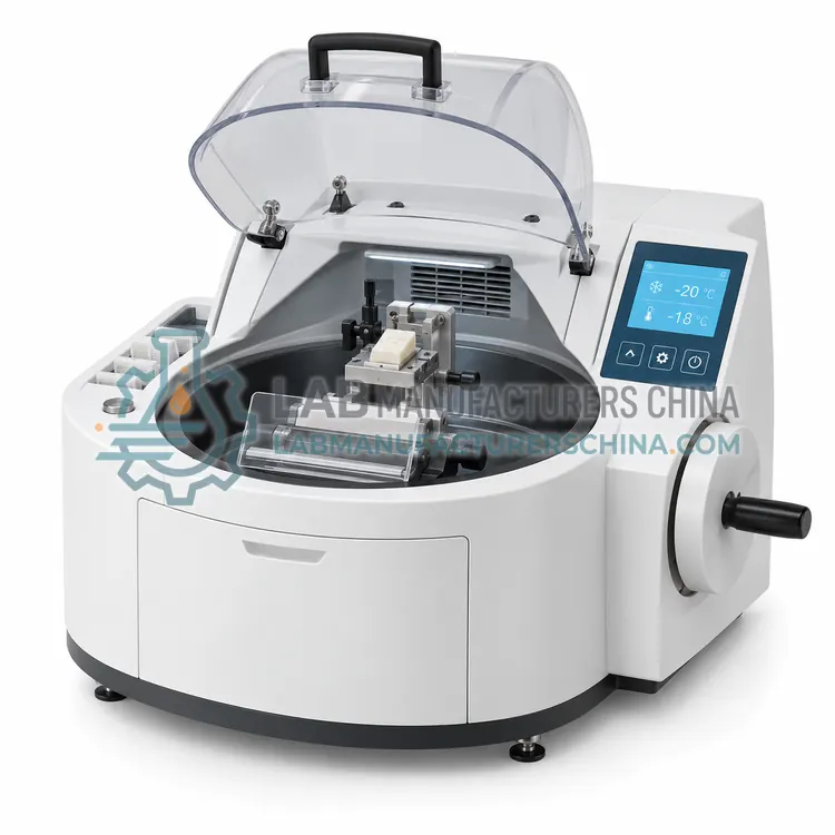
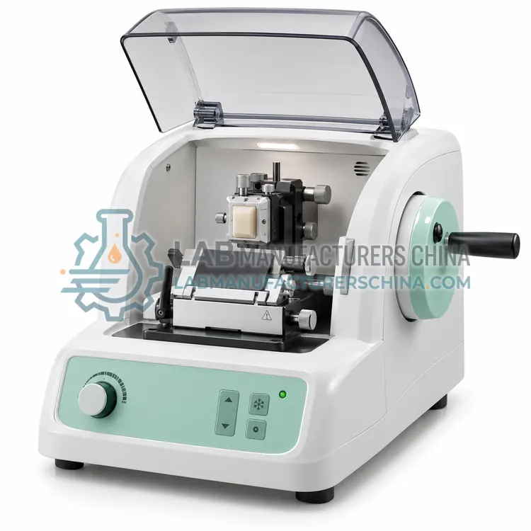
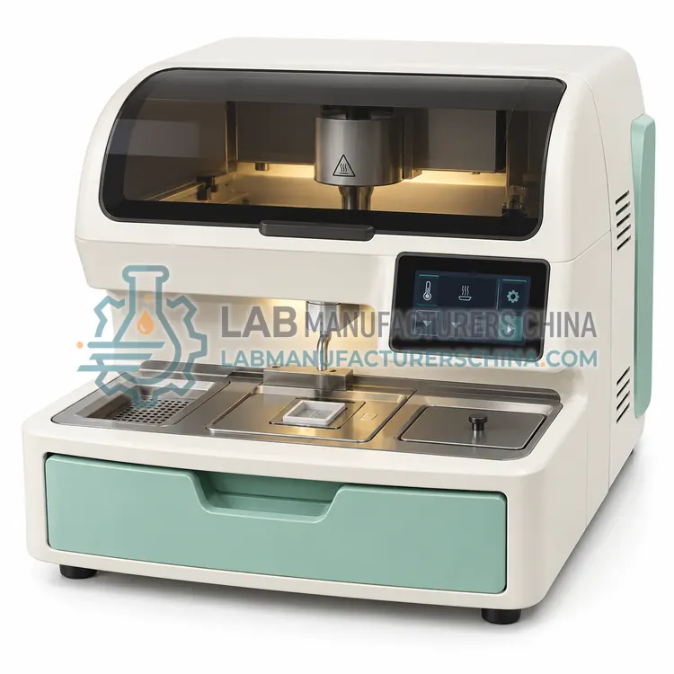
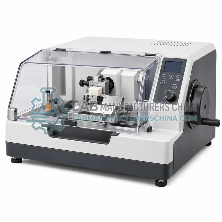
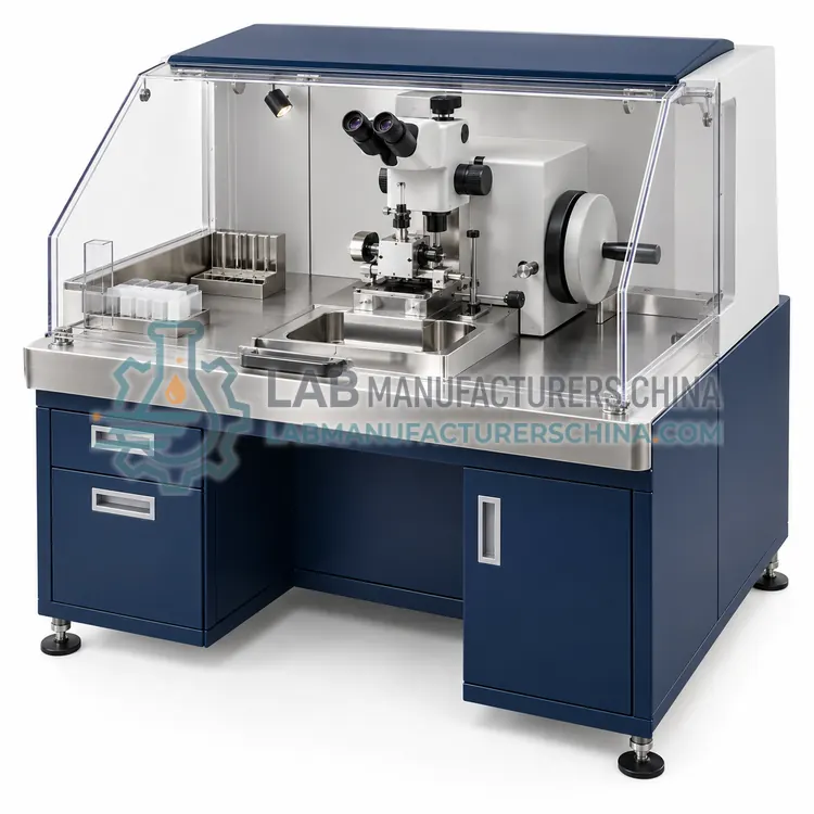
An ultramicrotome advances the specimen by nanometer-scale steps using thermal expansion of a rod or a precision mechanical feed, and cuts with a glass or diamond knife as the arm moves through a vertical stroke. Section thickness is set from 10 nm to 2 µm, with ultrathin sections of 40 to 100 nm used for TEM. Features include an illuminated stereomicroscope, knife and block approach controls, an automatic cutting cycle with adjustable speed from 0.1 to 10 mm/s, and an antistatic device. Cryo-ultramicrotomy is available with a cooled chamber for vitrified or frozen hydrated samples. Stable room conditions and vibration isolation are required, and operators need training to handle knives, troughs and grids.
How does it work?
A microtome holds a specimen block and advances it by a controlled increment toward a fixed knife or blade, then cuts a section as the block or knife moves past. Precision comes from a feed mechanism, often a micrometer screw or stepper motor, that sets section thickness, and from rigid, vibration-free mounting. Sliding microtomes move the block across a stationary knife on linear guides, while vibrating blade types oscillate the blade at 50 to 100 Hz to cut soft unfixed tissue. Ultramicrotomes use thermal or mechanical advance and a glass or diamond knife to cut 40 to 100 nm sections. Cooling chambers, knife angle adjustments, waste removal and safety guards are common components. Sharp, properly angled knives are essential for clean sections.
What are the key features?
- Thermal or mechanical advance with nanometer-scale steps
- Section thickness from 10 nm to 2 µm
- Automatic cutting cycle with adjustable speed
- Illuminated stereomicroscope for block and knife approach
- Antistatic device for handling sections
- Optional cryo chamber for frozen hydrated samples
What are the technical specifications?
A typical ultramicrotome configuration offers section thickness of 10 nm–2 µm, ultrathin setting of 40–100 nm, feed resolution of 1 nm, cutting speed of 0.1–10 mm/s, knife type of Glass or diamond knife and specimen block size of about 2 × 2 mm block face. Values are indicative of the product class; the exact figures for each configuration are confirmed on the quotation and the calibration documents supplied with the instrument.
Which applications is it used for?
The Ultramicrotome is used in transmission electron microscopy of tissue, virology and cell biology, materials science and polymer and composite analysis. Across the wider Microtomes class, laboratories also deploy it for surgical pathology, neuroscience research and electron microscopy.
Which standards and certifications apply?
Relevant standards and methods include IEC 61010-1, IEC 61010-2-101, IEC 61326-1, ISO 15189 and ISO 13485. Certification candidates for this model are CE, IEC 61010-1 compliance and ISO 13485 manufacturing; each is to be confirmed for the specific configuration before shipment, and certificates are supplied only where they have been issued.
Install the microtome on a rigid bench free of vibration and drafts, and keep it clean of debris. Handle blades and knives with extreme care, using guards and cut-resistant gloves, and dispose of used blades in a sharps container. Lubricate moving guides as recommended and check thickness calibration periodically with test sections or a calibrated gauge. Keep the block holder and knife clamp tight to prevent chatter. Ultramicrotomes need a stable room temperature and low air movement, while cryo models need regular defrosting and cleaning. Train staff on safe use and maintain records of service and calibration.
How do I select the right Ultramicrotome?
Select an ultramicrotome when sections must be under 200 nm for TEM. It requires a stable room, skilled users and regular knife costs, so it is not for routine histology. Specify feed resolution, cutting speed range, knife type, illumination, cryo option and any automation, and confirm service and training support.
How does it compare with other Microtomes variants?
Start with the specimen and the section thickness required. Paraffin blocks for routine histology use rotary models covered separately, while large, hard or frozen specimens call for sliding, freezing or saw microtomes. Electron microscopy requires an ultramicrotome, and fresh unfixed tissue for neuroscience suits a vibrating blade microtome. Compare thickness range and increment, specimen travel, knife type and holder, and whether motorized control is needed. Check stability, vibration isolation, ease of knife change, safety features and availability of consumables such as blades. Consider how many sections per day you will cut and whether cooling or chamber options are needed. Confirm service support and training.
Full specifications
| Section thickness | 10 nm–2 µm |
|---|---|
| Ultrathin setting | 40–100 nm |
| Feed resolution | 1 nm |
| Cutting speed | 0.1–10 mm/s |
| Knife type | Glass or diamond knife |
| Specimen block size | about 2 × 2 mm block face |
| Microscope magnification | 6–40× |
| Cryo option | to –160 °C |
| Power supply | 220 V, 50 Hz, about 0.3 kW |
Indicative values for this product class. Final specifications are confirmed on the quotation and in the calibration documents supplied with the instrument.
Applications
- Transmission electron microscopy of tissue
- Virology and cell biology
- Materials science
- Polymer and composite analysis
Key features
- Thermal or mechanical advance with nanometer-scale steps
- Section thickness from 10 nm to 2 µm
- Automatic cutting cycle with adjustable speed
- Illuminated stereomicroscope for block and knife approach
- Antistatic device for handling sections
- Optional cryo chamber for frozen hydrated samples
Frequently asked questions about Ultramicrotome
Glass knives made on a knife maker are used for trimming and preliminary sections, while diamond knives give consistent ultrathin sections of 40 to 100 nm and last for many thousands of sections. Diamond knives need careful cleaning and storage.
Air drafts, temperature swings and vibration cause thickness variation and chatter at nanometer scales. Place the instrument on an isolated bench in a stable room, away from doors, air vents and heavy equipment.
Cryo-ultramicrotomy cuts frozen hydrated or cryo-fixed samples at about –100 to –160 °C in a cooled chamber, preserving native structure without resin embedding. It needs cryo accessories, cooled knives and specially prepared grids.
A microtome cuts thin sections of tissue or material for microscopy. The sections can be a few nanometers to hundreds of micrometers thick, depending on the type, and are used in pathology, research, materials science and electron microscopy.
Rotary microtomes for paraffin work cut 1 to 60 µm, vibrating microtomes 20 to 500 µm, and ultramicrotomes 30 to 100 nm with glass or diamond knives. Actual results depend on the specimen, blade sharpness and stable installation.
A cryostat is a microtome inside a refrigerated chamber that cuts frozen tissue at about –20 °C. Other microtomes cut embedded or fixed specimens at room temperature, or fresh tissue in liquid in the case of vibrating models.
Related models