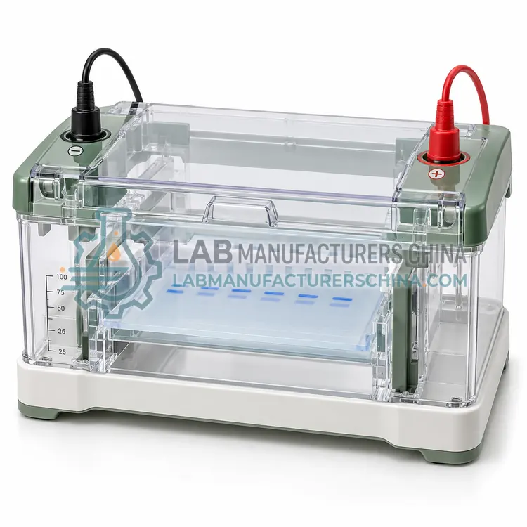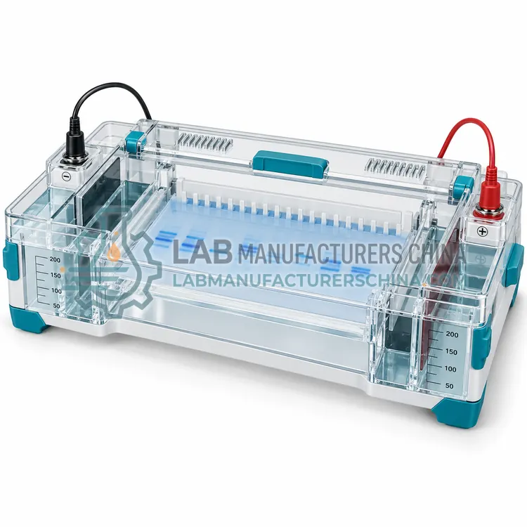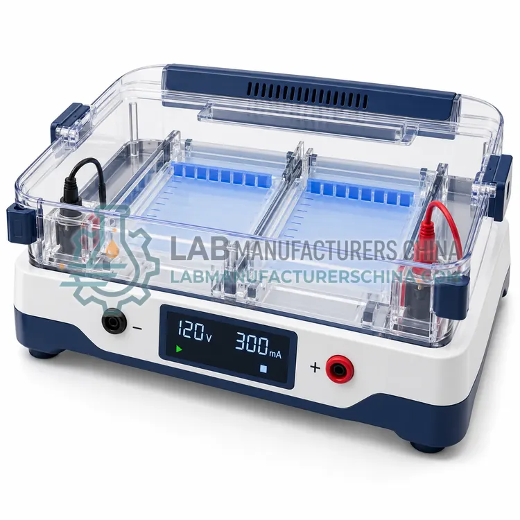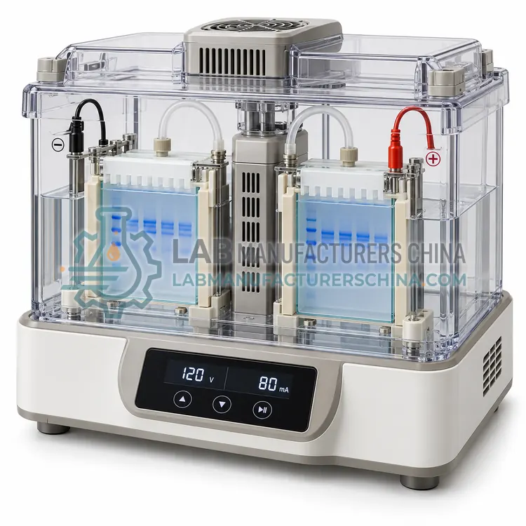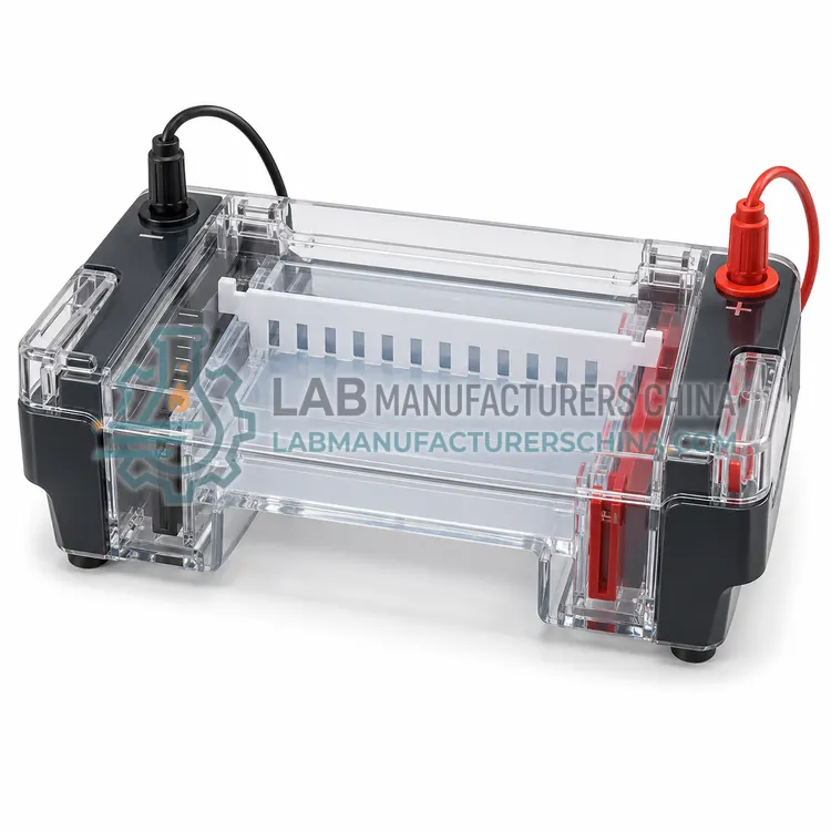
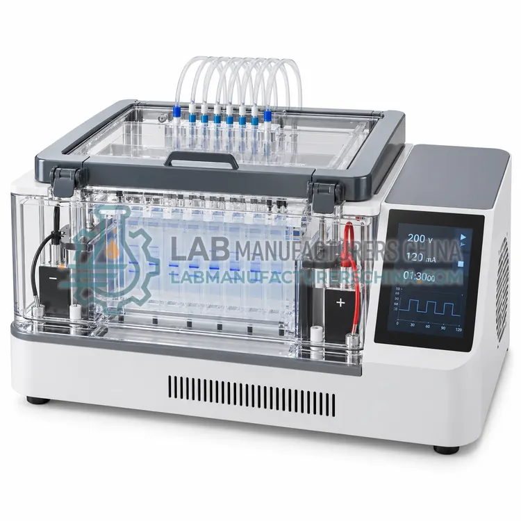
Pulsed-Field Gel Electrophoresis System
脉冲场凝胶电泳系统
- SKU HOE-0005
- Horizontal Electrophoresis
- CE (to be confirmed)
- ISO 9001 manufacturing (to be confirmed)
- RoHS (to be confirmed)
Price on request — FOB / CIF quoted per order
Prices depend on configuration, quantity and destination port.
Key specifications
| Separation range | approx. 10 kb to 5 Mb |
|---|---|
| Gel size | 14 x 13 cm or 20 x 20 cm |
| Field angle | 120° (CHEF hexagonal array) |
| Switch time range | 0.1 s – 18 min |
| Field strength | up to 6 V/cm |
| Buffer temperature control | approx. 10–20 °C |
Request a quote Download brochure · to be provided
By Lab Manufacturers China Technical TeamLast reviewed Standards: IEC 61010-1, IEC 61326-1, ISO 17025
What is the Pulsed-Field Gel Electrophoresis System?
A horizontal system that switches the electric field direction periodically to separate very large DNA molecules from about 10 kb up to several megabases. It is used for bacterial strain typing and large-fragment genome analysis.
What is the Pulsed-Field Gel Electrophoresis System?
A horizontal system that switches the electric field direction periodically to separate very large DNA molecules from about 10 kb up to several megabases. It is used for bacterial strain typing and large-fragment genome analysis.
Overview
Standard agarose gels cannot separate DNA above roughly 20–50 kb because large molecules migrate in a size-independent manner. Pulsed-field gel electrophoresis overcomes this by periodically changing the field direction, forcing molecules to reorient before they continue, with larger molecules taking longer. A typical system combines a cooled horizontal chamber, a hexagonal electrode array for contour-clamped homogeneous fields, a recirculating pump, a cooling module holding buffer at about 14 °C and a programmable controller. Switch times range from under a second to several minutes, and runs last 18–48 hours. Samples are embedded in agarose plugs to protect DNA from shearing. The technique is used for outbreak typing of Salmonella, Escherichia coli and Listeria, and for separating chromosomes of yeast and other organisms.
How does it work?
In a horizontal system the agarose gel is cast in a tray, placed in a tank and submerged a few millimetres below running buffer, usually TAE or TBE. When voltage is applied between the platinum or stainless-steel electrodes, negatively charged nucleic acids migrate toward the anode through the pores of the agarose. Shorter fragments move faster, so the bands sort by length, and a size ladder allows estimation of fragment size. The buffer carries current and conducts away Joule heat, which is why voltage per centimetre of electrode spacing is limited, typically to 5–10 V/cm. Bands are visualised with intercalating dyes such as ethidium bromide or safer alternatives under UV or blue light. Key components are the tank, gel tray, combs that form wells, electrode leads and a safety lid.
What are the key features?
- Contour-clamped homogeneous electric field electrode array gives straight lanes across the gel
- Programmable switch times from about 0.1 s to several minutes with ramped multi-block protocols
- Integrated cooling module and recirculation pump hold buffer near 14 °C during long runs
- Separates DNA from about 10 kb to several megabases using the same gel format
- Plug-casting accessories embed cells in agarose to protect large DNA from shearing
- Software stores run protocols and logs voltage, temperature and switch times for traceability
What are the technical specifications?
A typical pulsed-field gel electrophoresis system configuration offers separation range of approx. 10 kb to 5 Mb, gel size of 14 x 13 cm or 20 x 20 cm, field angle of 120° (CHEF hexagonal array), switch time range of 0.1 s – 18 min, field strength of up to 6 V/cm and buffer temperature control of approx. 10–20 °C. Values are indicative of the product class; the exact figures for each configuration are confirmed on the quotation and the calibration documents supplied with the instrument.
Which applications is it used for?
The Pulsed-Field Gel Electrophoresis System is used in bacterial outbreak strain typing, food-borne pathogen surveillance, yeast chromosome separation and large restriction fragment analysis. Across the wider Horizontal Electrophoresis class, laboratories also deploy it for PCR product verification, restriction digest analysis and RNA integrity checking.
Which standards and certifications apply?
Relevant standards and methods include IEC 61010-1, IEC 61326-1, ISO 17025 and Ph. Eur. 2.2.31. Certification candidates for this model are CE, ISO 9001 manufacturing and RoHS; each is to be confirmed for the specific configuration before shipment, and certificates are supplied only where they have been issued.
Place the tank on a level, vibration-free bench away from sinks and keep the power supply outside the splash zone. Replace running buffer regularly, because ion depletion and pH drift degrade resolution after repeated runs. Clean tanks with mild detergent and rinse with deionised water; avoid solvents that craze acrylic. Inspect electrodes for corrosion and check that lid interlocks and lead insulation remain intact. Handle ethidium bromide and UV sources under local chemical and optical safety rules, or switch to safer stains. Electrical safety should follow IEC 61010-1. Record voltage, time, gel percentage and buffer batch for traceability, and use ladders appropriate to the fragment range.
How do I select the right Pulsed-Field Gel Electrophoresis System?
Select pulsed-field only when you must resolve DNA above about 50 kb, for instance in PFGE-based surveillance; standard tanks are cheaper for smaller fragments. Specify the separation range, gel sizes, cooling capacity, switch-time programming options and whether plug moulds and a reference-standard protocol are supplied.
How does it compare with other Horizontal Electrophoresis variants?
Choose the gel size first, because it fixes run time, sample number and resolution. Mini tanks give fast checks of a few samples, midi and standard formats balance throughput and resolution, and wide-format tanks accommodate multi-channel pipetting for 96-well workflows. Check the maximum voltage and buffer volume, as a larger tank needs a bigger power supply and more buffer. Consider whether dual trays or removable casting systems help speed. Pulsed-field systems are only needed for fragments above roughly 50 kb, such as genomic DNA typing. Also confirm comb thickness and tooth options, electrode material, lid interlock design and chemical resistance of the polycarbonate or acrylic body, and whether replacement trays and combs are easy to source.
Full specifications
| Separation range | approx. 10 kb to 5 Mb |
|---|---|
| Gel size | 14 x 13 cm or 20 x 20 cm |
| Field angle | 120° (CHEF hexagonal array) |
| Switch time range | 0.1 s – 18 min |
| Field strength | up to 6 V/cm |
| Buffer temperature control | approx. 10–20 °C |
| Buffer volume | approx. 2.0–2.5 L |
| Typical run time | 18–48 h |
| Recirculation pump flow | approx. 0.75–1.0 L/min |
| Power requirement | 100–240 V AC, 50/60 Hz |
Indicative values for this product class. Final specifications are confirmed on the quotation and in the calibration documents supplied with the instrument.
Applications
- Bacterial outbreak strain typing
- Food-borne pathogen surveillance
- Yeast chromosome separation
- Large restriction fragment analysis
Key features
- Contour-clamped homogeneous electric field electrode array gives straight lanes across the gel
- Programmable switch times from about 0.1 s to several minutes with ramped multi-block protocols
- Integrated cooling module and recirculation pump hold buffer near 14 °C during long runs
- Separates DNA from about 10 kb to several megabases using the same gel format
- Plug-casting accessories embed cells in agarose to protect large DNA from shearing
- Software stores run protocols and logs voltage, temperature and switch times for traceability
Frequently asked questions about Pulsed-Field Gel Electrophoresis System
In a constant field, DNA above roughly 20–50 kb stretches into the gel pores and migrates at a nearly size-independent rate. Periodic changes of field direction force each molecule to reorient, and larger molecules reorient more slowly, which restores size-dependent separation.
Megabase DNA is easily sheared by pipetting and vortexing. Embedding intact cells in agarose plugs and lysing them in place keeps the DNA whole, and restriction digestion can then be done inside the plug before loading into the gel.
Long runs at 18–48 hours would heat the buffer and distort migration or soften the gel. A chiller and recirculating pump hold the temperature near 14 °C, keeping buffer composition uniform and band positions reproducible between runs.
A horizontal electrophoresis system is a tank in which an agarose gel lies flat and submerged in buffer while voltage moves DNA or RNA fragments through it. Fragments separate by length, so laboratories use it to check PCR products, digests and nucleic acid quality.
A common starting point is 5–8 V per centimetre of distance between electrodes. Higher voltage shortens the run but raises heat and can smear bands or melt the gel. Lower voltage over a longer time gives sharper resolution of large fragments.
Replace running buffer after several runs, or sooner for long or high-voltage runs, because buffering capacity falls and pH drifts. Discoloured buffer, slow migration or distorted bands are signs it should be changed. Never top up with water.
Related models