
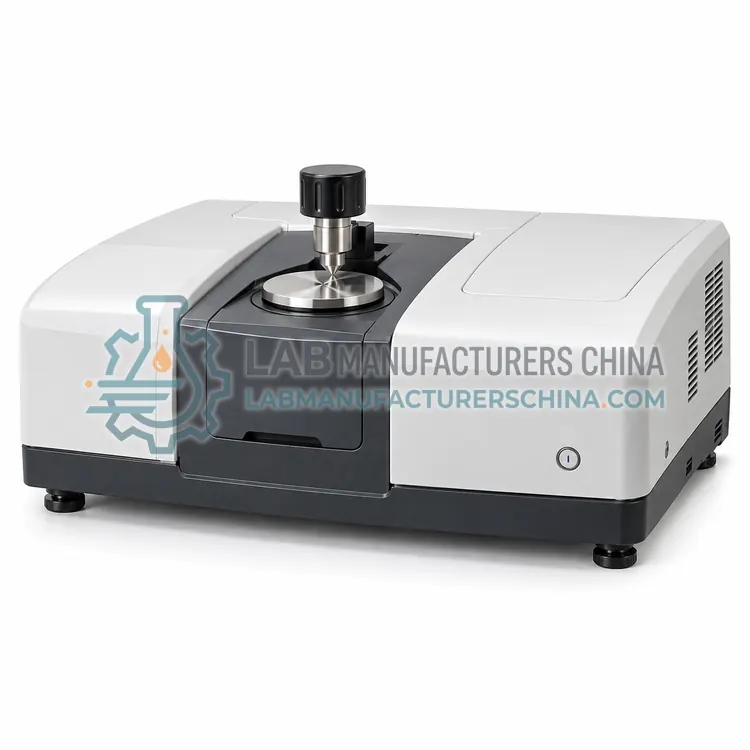
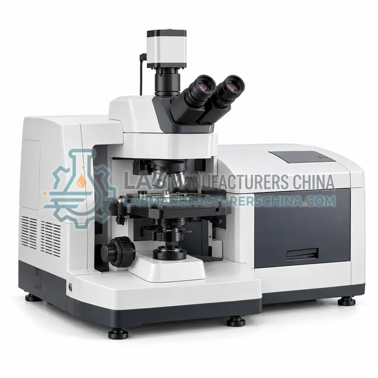
FTIR Microscope for Microanalysis and Chemical Imaging
用于微区分析和化学成像的红外显微镜
- SKU FTI-0003
- FTIR
- CE (to be confirmed)
- ISO 9001 manufacturing (to be confirmed)
- IEC 61010-1 safety (to be confirmed)
Price on request — FOB / CIF quoted per order
Prices depend on configuration, quantity and destination port.
Key specifications
| Spectral range | 4000–650 cm-1 (MCT) |
|---|---|
| Minimum sample area | 10 × 10 µm |
| Objectives | 15× and 36× Cassegrain, Ge ATR 20× |
| Detector | MCT (LN2) single element; FPA 64×64 or 128×128 option |
| Measurement modes | Transmission, reflection, ATR |
| Stage travel | ≥75 × 50 mm, 0.1 µm step |
Request a quote Download brochure · to be provided
By Lab Manufacturers China Technical TeamLast reviewed Standards: ASTM E1421, ASTM E168, ASTM E1252
What is the FTIR Microscope for Microanalysis and Chemical Imaging?
An FTIR microscope couples an infrared spectrometer to a visible and infrared microscope to analyze samples down to about 10 µm. It supports point analysis, line scans and chemical imaging with focal plane array detectors for contaminants, fibers, laminates and biological tissue.
What is the FTIR Microscope for Microanalysis and Chemical Imaging?
An FTIR microscope couples an infrared spectrometer to a visible and infrared microscope to analyze samples down to about 10 µm. It supports point analysis, line scans and chemical imaging with focal plane array detectors for contaminants, fibers, laminates and biological tissue.
Overview
The FTIR microscope uses reflective Cassegrain optics with 15× and 36× objectives, a motorized stage and aperture system to select areas as small as 10 × 10 µm. A liquid-nitrogen-cooled MCT detector provides the sensitivity needed for small samples, and operation modes include transmission, reflection and ATR using a germanium crystal objective for contact measurements at higher spatial resolution. A focal plane array of 64 × 64 or 128 × 128 elements images thousands of spectra in a single acquisition, forming chemical maps of polymer layers, particle distributions and tissue sections. Visible camera and motorized stage software locate features and automate mapping. Because diffraction limits resolution to approximately the wavelength, 3–10 µm, samples below about 10 µm cannot be resolved. It is widely used for microplastics, forensic fiber comparison and failure analysis of multilayer packaging.
How does it work?
An FTIR spectrometer directs broadband infrared radiation from a globar or ceramic source into a Michelson interferometer, where a beam splitter divides it between a fixed mirror and a moving mirror. The recombined beam produces an interferogram that varies with mirror displacement. The interferogram passes through or reflects from the sample, is recorded by a DTGS or cooled MCT detector, and is converted to a spectrum by a Fourier transform. Multiplexing allows all frequencies to be measured simultaneously, giving the Fellgett advantage, and the circular aperture gives the Jacquinot advantage in throughput. A helium-neon laser tracks mirror position to give wavenumber accuracy of 0.01 cm-1. Sampling accessories include attenuated total reflection crystals, transmission holders, diffuse reflectance cells and gas cells, allowing solids, liquids, films and gases to be analyzed with minimal preparation.
What are the key features?
- Reflective Cassegrain optics analyze samples down to 10 µm without chromatic errors.
- Transmission, reflection and ATR modes cover films, particles and thick samples.
- Focal plane array detector images thousands of spectra per acquisition.
- Motorized stage and visible camera automate mapping and area selection.
- Germanium ATR objective increases spatial resolution for contact analysis.
- Software creates chemical images from band height, area or ratios.
What are the technical specifications?
A typical ftir microscope for microanalysis and chemical imaging configuration offers spectral range of 4000–650 cm-1 (MCT), minimum sample area of 10 × 10 µm, objectives of 15× and 36× Cassegrain, Ge ATR 20×, detector of MCT (LN2) single element; FPA 64×64 or 128×128 option, measurement modes of Transmission, reflection, ATR and stage travel of ≥75 × 50 mm, 0.1 µm step. Values are indicative of the product class; the exact figures for each configuration are confirmed on the quotation and the calibration documents supplied with the instrument.
Which applications is it used for?
The FTIR Microscope for Microanalysis and Chemical Imaging is used in microplastics identification, forensic fiber and paint analysis, laminate and packaging failure analysis, pharmaceutical tablet and inclusion mapping and biological tissue histochemistry. Across the wider FTIR class, laboratories also deploy it for polymer and plastics identification, forensic trace evidence analysis and contaminant and failure analysis.
Which standards and certifications apply?
Relevant standards and methods include ASTM E1421, ASTM E168, ASTM E1252, ISO 10640, USP <854>, Ph. Eur. 2.2.24 and GB/T 6040. Certification candidates for this model are CE, ISO 9001 manufacturing and IEC 61010-1 safety; each is to be confirmed for the specific configuration before shipment, and certificates are supplied only where they have been issued.
Place the spectrometer on a stable, vibration-free bench with controlled humidity, since KBr optics are hygroscopic and water vapor interferes with spectra. Keep desiccant in the interferometer compartment or use a dry air or nitrogen purge. Collect a fresh background before each sample and clean the ATR crystal with isopropanol or a suitable solvent between measurements. Verify wavenumber accuracy with a polystyrene film and photometric performance with standard checks such as signal-to-noise and energy throughput. Replace the IR source and desiccant cartridges as recommended. Laser and MCT detector cooling with liquid nitrogen require trained handling. For regulated use, implement IQ/OQ/PQ and software with audit trails.
How do I select the right FTIR Microscope for Microanalysis and Chemical Imaging?
Select an FTIR microscope when samples or features are below roughly 500 µm or spatial distribution is important. For bulk material identification, ATR-FTIR is faster and less costly. In an RFQ, specify minimum area, objectives, detector type including FPA, stage size, measurement modes and software for chemical imaging.
How does it compare with other FTIR variants?
Choose an FTIR instrument by spectral resolution, sensitivity, sampling method and intended environment. Routine identification of polymers and organics is well served by a benchtop unit with 4 cm-1 resolution, a DTGS detector and a diamond ATR accessory. Research requiring 0.1 cm-1 resolution, step-scan time resolution or far-IR coverage needs a research-grade interferometer with optional MCT detectors. Microscopic contaminants below 50 µm require an FTIR microscope with mapping. Field identification of unknown chemicals favors a ruggedized handheld unit, and emissions or atmospheric work needs a gas analyzer with a heated multipass cell. Evaluate signal-to-noise ratio, library availability, purge requirements, accessory flexibility and software features for regulated laboratories.
Full specifications
| Spectral range | 4000–650 cm-1 (MCT) |
|---|---|
| Minimum sample area | 10 × 10 µm |
| Objectives | 15× and 36× Cassegrain, Ge ATR 20× |
| Detector | MCT (LN2) single element; FPA 64×64 or 128×128 option |
| Measurement modes | Transmission, reflection, ATR |
| Stage travel | ≥75 × 50 mm, 0.1 µm step |
| Mapping speed (FPA) | 4096 spectra in <1 min |
| Spectral resolution | 4 cm-1 (2 cm-1 optional) |
| Visible imaging | Color camera, 1–10 MP |
| Coupled spectrometer | Research or benchtop FTIR |
Indicative values for this product class. Final specifications are confirmed on the quotation and in the calibration documents supplied with the instrument.
Applications
- Microplastics identification
- Forensic fiber and paint analysis
- Laminate and packaging failure analysis
- Pharmaceutical tablet and inclusion mapping
- Biological tissue histochemistry
Key features
- Reflective Cassegrain optics analyze samples down to 10 µm without chromatic errors.
- Transmission, reflection and ATR modes cover films, particles and thick samples.
- Focal plane array detector images thousands of spectra per acquisition.
- Motorized stage and visible camera automate mapping and area selection.
- Germanium ATR objective increases spatial resolution for contact analysis.
- Software creates chemical images from band height, area or ratios.
Frequently asked questions about FTIR Microscope for Microanalysis and Chemical Imaging
Diffraction limits mid-infrared spatial resolution to roughly the wavelength, so practical minimum sizes are about 10 µm in transmission or reflection and 3–5 µm with germanium ATR. Smaller features give mixed or distorted spectra.
An FPA is a two-dimensional detector, for example 64 × 64 pixels, that records an infrared spectrum at each pixel simultaneously. It produces chemical images in minutes instead of hours required by mapping with a single-element detector.
Yes. FTIR microscopy in reflection, transmission or ATR can identify polymer types of particles typically larger than 10–20 µm by library matching. Automated particle finding and FPA imaging speed up analysis of filter samples.
FTIR spectroscopy identifies materials from their infrared absorption fingerprint. It is used to determine functional groups, identify polymers and contaminants, confirm raw materials, study coatings and films and quantify components in mixtures or gases without lengthy sample preparation.
Attenuated total reflection passes the infrared beam through a high-refractive-index crystal such as diamond. An evanescent wave penetrates a few micrometers into the sample pressed on the crystal, so solids, pastes and liquids can be measured with almost no preparation.
A resolution of 4 cm-1 is sufficient for most condensed-phase identification, with 2 cm-1 used for some library matching. Gas-phase work and rotational fine structure need 0.5 cm-1 or better, which requires a research-grade interferometer.
Related models
UV-Vis Spectrophotometer with Peltier Multi-Cell Kinetics System
带帕尔贴多池动力学系统的紫外可见分光光度计
- CE (to be confirmed)
- +1