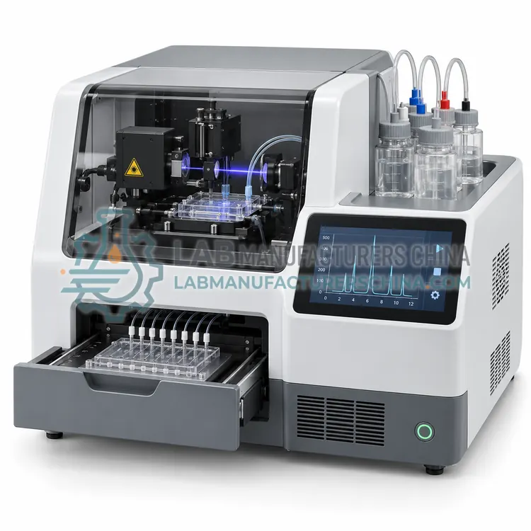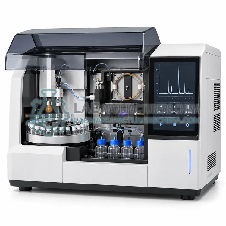
Capillary Zone Electrophoresis System with UV-DAD Detection
配紫外二极管阵列检测器的毛细管区带电泳系统
- CE (to be confirmed)
- +2
Welcome to Lab Manufacturers China — laboratory instruments direct from a Chinese manufacturer
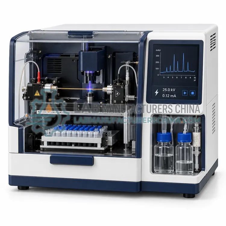
毛细管等电聚焦(cIEF)系统
Price on request — FOB / CIF quoted per order
Prices depend on configuration, quantity and destination port.
| Detection | Whole-column UV imaging at 280 nm (or single-point UV) |
|---|---|
| Capillary coating | Neutral (EOF-suppressed) |
| Capillary internal diameter | 50–100 µm |
| Capillary length | approx. 5–10 cm (imaging) or 30 cm |
| Focusing voltage | up to 30 kV |
| pI resolution | approx. 0.05 pI units |
By Lab Manufacturers China Technical TeamLast reviewed Standards: USP <1053>, Ph. Eur. 2.2.47, IEC 61010-1
A capillary instrument that focuses proteins along a pH gradient formed by carrier ampholytes and images or detects them by isoelectric point, used to profile charge variants of antibodies and recombinant proteins.
A capillary instrument that focuses proteins along a pH gradient formed by carrier ampholytes and images or detects them by isoelectric point, used to profile charge variants of antibodies and recombinant proteins.
Capillary isoelectric focusing separates proteins by isoelectric point in a pH gradient created by carrier ampholytes inside a neutral-coated capillary. Sample is mixed with ampholytes and pI markers, loaded, and focused at 15–30 kV across the capillary, typically for 5–10 minutes. In imaging systems the focused zones are detected along the whole capillary at 280 nm without mobilisation, which gives quick, reproducible profiles, while in conventional cIEF the zones are chemically or hydrodynamically mobilised past a detector. The technique resolves charge variants such as deamidated, sialylated and C-terminal lysine forms to about 0.05 pI units, and pI markers convert the profile to a pH scale. It is used for release and stability testing of monoclonal antibodies and other biologics, where charge heterogeneity is a critical quality attribute.
In capillary electrophoresis a fused-silica capillary of 25–100 micrometres internal diameter is filled with a background electrolyte or sieving polymer and connected between two buffer vials with electrodes. A few nanolitres of sample are injected hydrodynamically or electrokinetically and 5–30 kV is applied. The narrow bore dissipates heat efficiently, so high fields can be used, giving fast and sharp separations. Ions migrate according to their charge-to-size ratio, and in capillary zone electrophoresis electroosmotic flow from the charged silica wall carries all species toward the detector. Gel or polymer sieving separates DNA and SDS-protein complexes by size, while isoelectric focusing resolves proteins by pI. Detection is by UV absorbance or diode array, laser-induced fluorescence or mass spectrometry, and data are processed as electropherograms.
A typical capillary isoelectric focusing (cief) system configuration offers detection of Whole-column UV imaging at 280 nm (or single-point UV), capillary coating of Neutral (EOF-suppressed), capillary internal diameter of 50–100 µm, capillary length of approx. 5–10 cm (imaging) or 30 cm, focusing voltage of up to 30 kV and pI resolution of approx. 0.05 pI units. Values are indicative of the product class; the exact figures for each configuration are confirmed on the quotation and the calibration documents supplied with the instrument.
The Capillary Isoelectric Focusing (cIEF) System is used in monoclonal antibody charge variant profiling, biosimilar comparability studies, stability and forced-degradation studies and pI determination of recombinant proteins. Across the wider Capillary Electrophoresis class, laboratories also deploy it for monoclonal antibody purity and charge variants, STR profiling for forensic identity and DNA fragment sizing and genotyping.
Relevant standards and methods include USP <1053>, Ph. Eur. 2.2.47, IEC 61010-1, IEC 60825-1, 21 CFR Part 11 and ISO 18385. Certification candidates for this model are CE, ISO 9001 manufacturing and 21 CFR Part 11 software compliance; each is to be confirmed for the specific configuration before shipment, and certificates are supplied only where they have been issued.
Install the instrument on a stable bench with clean power, controlled ambient temperature and, for fluorescence systems, stable laser and optics conditions. Condition new capillaries according to the method, and flush between runs to maintain reproducible electroosmotic flow. Use filtered, degassed buffers and keep vials free of particles, since blockages and bubbles are the most common faults. Replace capillaries, polymer and windows on schedule, and verify performance with size standards or system suitability mixtures. Regulated users should plan IQ/OQ/PQ, calibration and software validation, with audit trails under 21 CFR Part 11. Safety follows IEC 61010-1 with interlocked high-voltage compartments, and laser safety under IEC 60825-1 applies to fluorescence detectors.
Select a cIEF system for charge-variant testing of biologics where gel IEF is too slow or qualitative. Specify imaging or mobilisation detection, capillary coating and dimensions, pH range, sample capacity and availability of validated method kits and compliance documentation.
Choose by application first. Capillary zone electrophoresis with UV or diode-array detection covers small ions, peptides, drugs and charge variants. Multi-capillary array analyzers with laser-induced fluorescence suit STR typing, fragment analysis and sequencing. Gel-based CE-SDS is the standard for size purity of monoclonal antibodies, and capillary isoelectric focusing resolves charge heterogeneity. Microchip formats trade resolution for speed and low sample use. Compare capillary count, detection type and sensitivity, temperature control, injection repeatability, autosampler capacity, and the software's audit trail and validation package for regulated use. Also consider consumable cost, such as capillaries, polymer and buffers, and whether the supplier can provide IQ/OQ documentation and method transfer support.
| Detection | Whole-column UV imaging at 280 nm (or single-point UV) |
|---|---|
| Capillary coating | Neutral (EOF-suppressed) |
| Capillary internal diameter | 50–100 µm |
| Capillary length | approx. 5–10 cm (imaging) or 30 cm |
| Focusing voltage | up to 30 kV |
| pI resolution | approx. 0.05 pI units |
| pH gradient range | pH 3–10 (ampholyte-dependent) |
| Focusing time | approx. 5–10 min |
| Temperature control | approx. 10–25 °C |
| Sample capacity | 48–96 samples |
Indicative values for this product class. Final specifications are confirmed on the quotation and in the calibration documents supplied with the instrument.
cIEF measures the distribution of protein forms by isoelectric point, showing acidic, main and basic charge variants. It is used to characterise monoclonal antibodies, where modifications such as deamidation, sialylation or lysine truncation change net charge.
Coating removes the charged silanol groups that generate electroosmotic flow. Without it, flow would disturb the focused zones, so a neutral coating lets proteins settle at their isoelectric points and stay sharply focused.
Imaged cIEF detects all focused zones across the whole capillary at once, so no mobilisation step is needed, which shortens analysis and improves reproducibility. Conventional cIEF mobilises zones past a single detection point and can distort the profile.
Capillary electrophoresis separates ions, proteins and nucleic acids in a narrow capillary with on-line detection. It is used for antibody purity and charge variant analysis, STR profiling, DNA fragment sizing, sequencing and the analysis of small ions, drugs and organic acids.
CE separates by charge-to-size ratio in an electric field using tiny sample volumes and no pumped mobile phase, giving high efficiency and low solvent use. HPLC separates by partitioning between mobile and stationary phases and handles larger volumes and neutral analytes more easily.
CE-SDS is capillary gel electrophoresis of SDS-denatured proteins in a sieving polymer. It separates fragments by molecular weight and is widely used to measure size purity of monoclonal antibodies, reduced and non-reduced, as a replacement for slab SDS-PAGE in quality control.
配紫外二极管阵列检测器的毛细管区带电泳系统
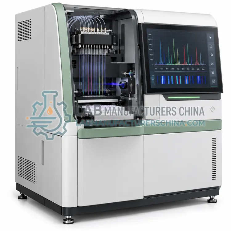
配激光诱导荧光的多毛细管阵列遗传分析仪
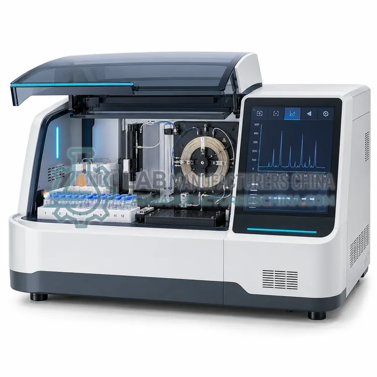
用于 CE-SDS 蛋白分析的毛细管凝胶电泳系统