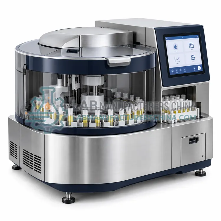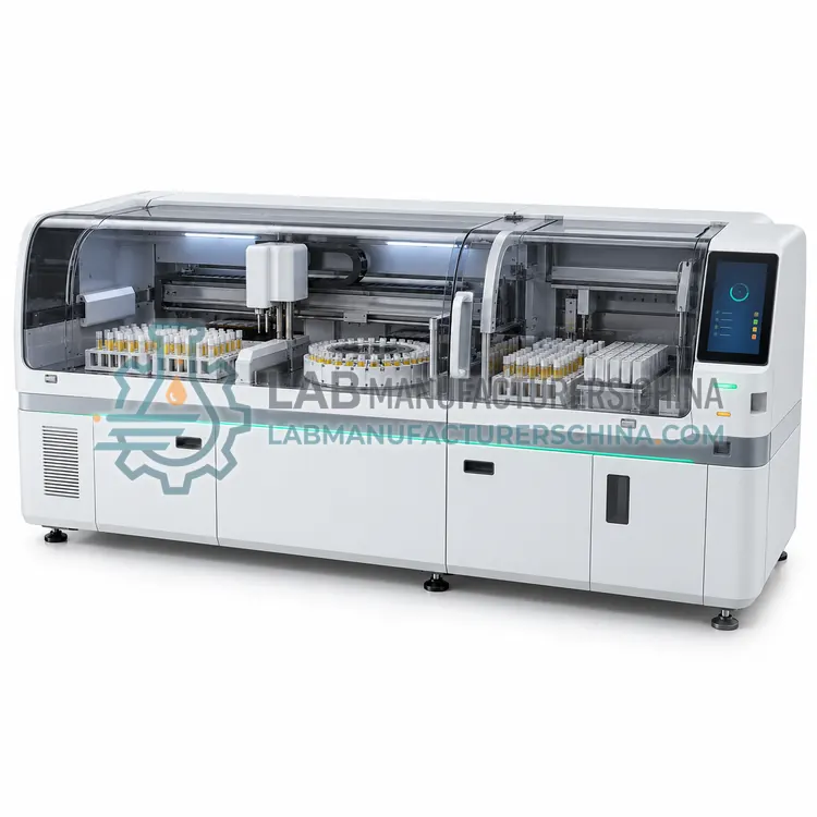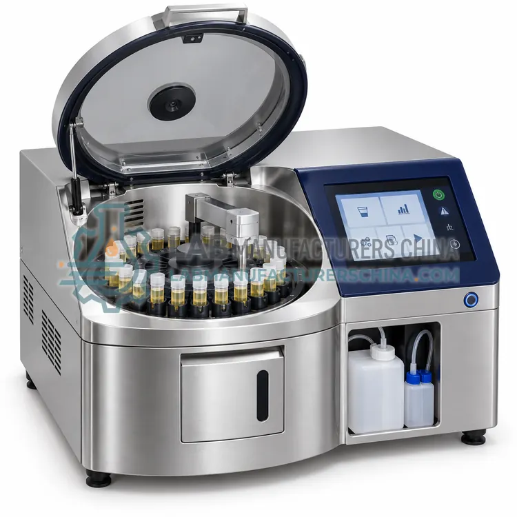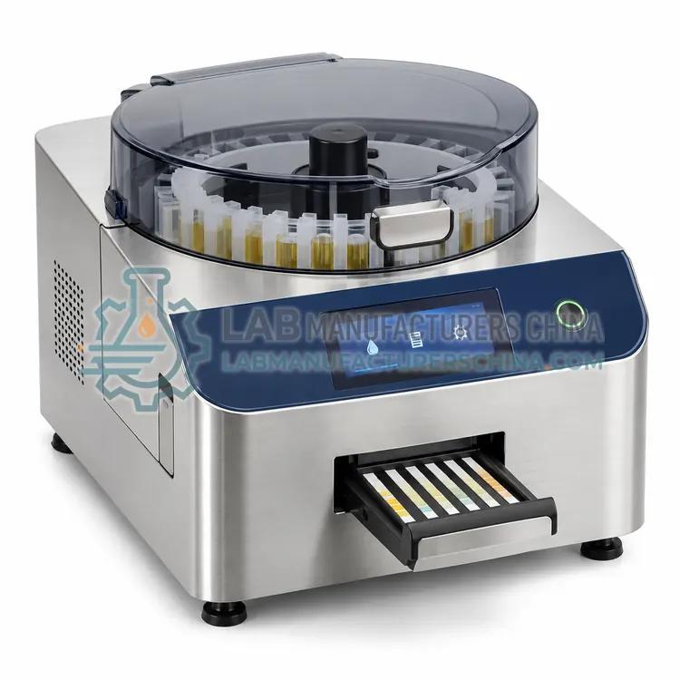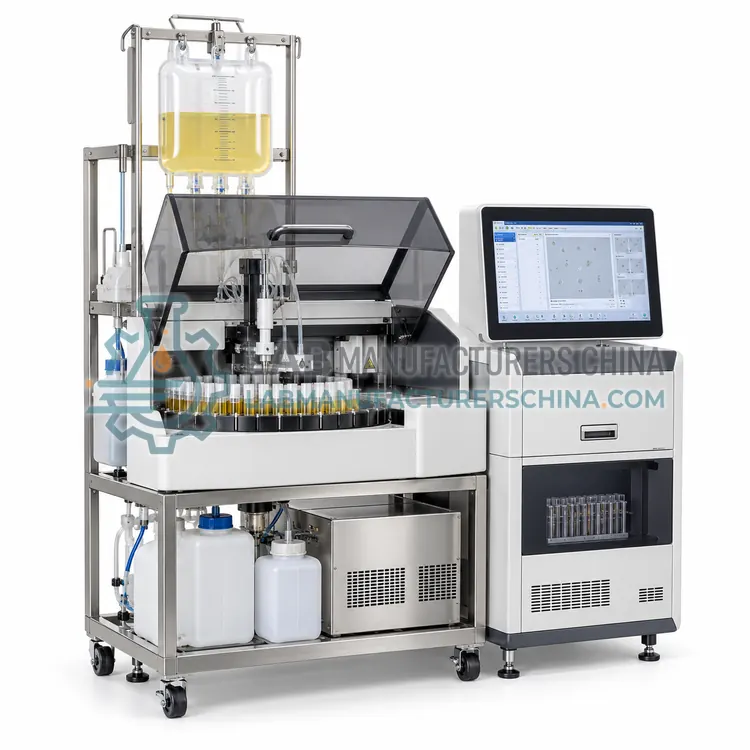
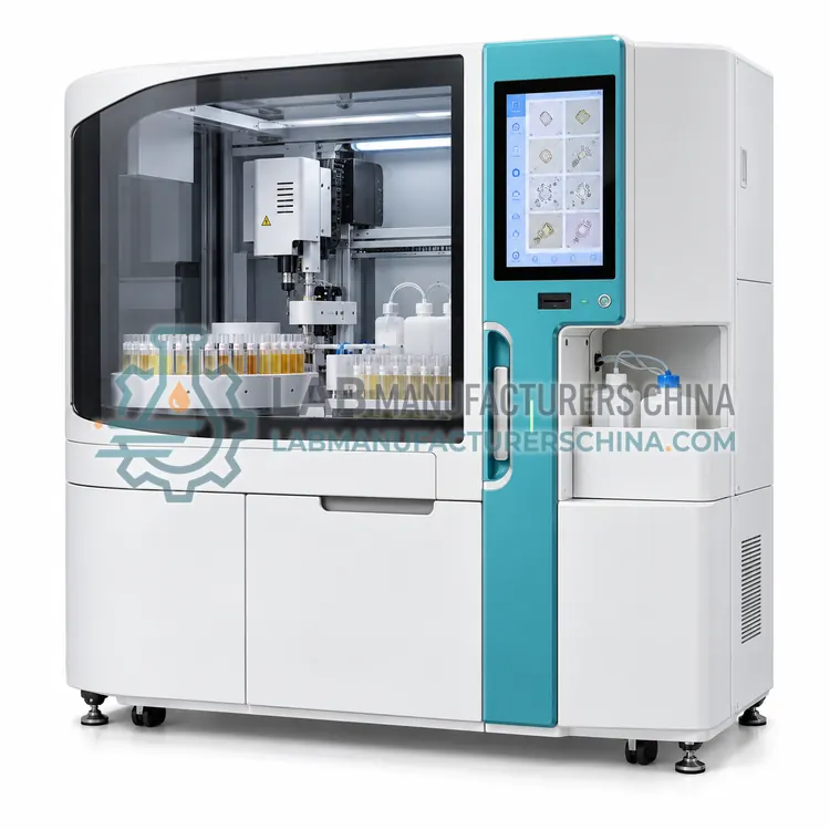
Fully Automated Urinalysis Analyzer with Flow Imaging Sediment Module
带流动成像沉渣模块的全自动尿液分析仪
- SKU AUS-0002
- Automated Urinalysis Systems
- CE (to be confirmed)
- ISO 13485 manufacturing (to be confirmed)
- NMPA class II registration (to be confirmed)
Price on request — FOB / CIF quoted per order
Prices depend on configuration, quantity and destination port.
Key specifications
| Sediment method | Flow imaging with digital pattern recognition |
|---|---|
| Images per sample | Up to 1,000 particle images stored |
| Particle classes | RBC, WBC, epithelial, casts, crystals, bacteria, yeast |
| Throughput | 60–120 samples/h combined |
| Sample volume | 1–2 mL per sediment analysis |
| Image resolution | About 1 µm per pixel |
Request a quote Download brochure · to be provided
By Lab Manufacturers China Technical TeamLast reviewed Standards: ISO 15189, CLSI GP16, CLSI EP05
What is the Fully Automated Urinalysis Analyzer with Flow Imaging Sediment Module?
This analyzer uses flow imaging in the sediment module, capturing digital images of particles as they pass a camera and classifying them with pattern recognition. It retains images for review, which suits laboratories that want visual evidence with each sediment result.
What is the Fully Automated Urinalysis Analyzer with Flow Imaging Sediment Module?
This analyzer uses flow imaging in the sediment module, capturing digital images of particles as they pass a camera and classifying them with pattern recognition. It retains images for review, which suits laboratories that want visual evidence with each sediment result.
Overview
In flow imaging, a diluted urine sample passes through a thin flow cell, and a camera takes images of each particle as it moves through the field of view. Software segments the images and classifies particles such as red cells, white cells, epithelial cells, casts, crystals, bacteria and yeast, reporting counts per volume. Because the original images are stored and linked to the result, a technologist can verify borderline classifications on screen without preparing a slide, and atypical findings can be reviewed later. The instrument also measures particle size and can flag images that do not match any stored class, which helps detect unusual elements. Image quality depends on correct dilution and on cleaning of the flow cell, since deposits appear as artefacts in the images.
How does it work?
The platform links two analytical modules through one sample and one worklist. The chemistry module dips a reagent strip, reads the pads by reflectance photometry at defined wavelengths, applies colour and ascorbate compensation and reports semiquantitative categories. The sediment module either uses flow cytometry with a nucleic acid stain and scatter signals to classify particles by size, granularity and fluorescence, or images a sediment chamber or centrifuged slide and classifies particles by pattern recognition. The software merges both result sets into one report, applies review rules such as reflex microscopy for flagged samples, and transmits results to the laboratory information system. Sample barcode reading, mixing before aspiration and automatic washing between samples reduce operator handling and improve traceability, while quality control records and reagent lot tracking support accreditation.
What are the key features?
- Camera-based flow imaging of particles in a thin flow cell
- Pattern recognition classification with per-volume counts
- Stored images linked to each sediment result
- On-screen verification of borderline classifications
- Flags for images not matching stored particle classes
- Particle size measurement reported with counts
What are the technical specifications?
A typical fully automated urinalysis analyzer with flow imaging sediment module configuration offers sediment method of Flow imaging with digital pattern recognition, images per sample of Up to 1,000 particle images stored, particle classes of RBC, WBC, epithelial, casts, crystals, bacteria, yeast, throughput of 60–120 samples/h combined, sample volume of 1–2 mL per sediment analysis and image resolution of About 1 µm per pixel. Values are indicative of the product class; the exact figures for each configuration are confirmed on the quotation and the calibration documents supplied with the instrument.
Which applications is it used for?
The Fully Automated Urinalysis Analyzer with Flow Imaging Sediment Module is used in urinalysis with image documentation, teaching and competency assessment of sediment findings, nephrology sample review with archived evidence and laboratories auditing classification accuracy. Across the wider Automated Urinalysis Systems class, laboratories also deploy it for hospital routine urinalysis with combined reporting, nephrology and urology outpatient testing and emergency department urine screening.
Which standards and certifications apply?
Relevant standards and methods include ISO 15189, CLSI GP16, CLSI EP05, CLSI AUTO10, CLSI POCT04, IEC 61010-1 and IEC 61010-2-101. Certification candidates for this model are CE, ISO 13485 manufacturing and NMPA class II registration; each is to be confirmed for the specific configuration before shipment, and certificates are supplied only where they have been issued.
Install the system on a level bench with controlled temperature, stable power and enough space for reagent and waste containers, and verify each module separately before validating the combined workflow with a set of normal and abnormal samples compared against manual microscopy. Define the review rules and test them before go-live, since rule errors affect which results are released automatically. Daily work includes running chemistry control strips and sediment control material, checking reagent and strip status and inspecting the flow cell or imaging chamber for deposits. Wash solutions, cuvettes and flow cells require scheduled cleaning, and results flagged by the system should be reviewed according to documented criteria. Treat all urine samples and waste as potentially infectious.
How do I select the right Fully Automated Urinalysis Analyzer with Flow Imaging Sediment Module?
Specify flow imaging when archived images and on-screen verification are valuable, for example for teaching, audit or when clinicians request supporting evidence. In the RFQ confirm storage requirements, image export formats, the classification classes supported and the volume of data generated per sample.
How does it compare with other Automated Urinalysis Systems variants?
Choose an automated urinalysis system when sediment results are required routinely and the daily sample number exceeds a few hundred, or when manual microscopy cannot keep up with the workload. Compare systems by which sediment technology is used, the particle classes reported, whether a microscope image is available for review, and the review rules offered. Check the chemistry parameters, cross-contamination control between samples, barcode handling and the combined report format. Confirm the throughput on a realistic mixture of normal and abnormal samples, since flagged samples take longer, and the sample volume needed for both modules. Verify LIS integration and the traceability features, and plan a downtime procedure because a single platform carries both chemistry and sediment reporting.
Full specifications
| Sediment method | Flow imaging with digital pattern recognition |
|---|---|
| Images per sample | Up to 1,000 particle images stored |
| Particle classes | RBC, WBC, epithelial, casts, crystals, bacteria, yeast |
| Throughput | 60–120 samples/h combined |
| Sample volume | 1–2 mL per sediment analysis |
| Image resolution | About 1 µm per pixel |
| Data storage | Images archived on server or local storage |
| Interfaces | LAN with HL7 or ASTM, image export option |
| Power supply | AC 100–240 V, 50/60 Hz |
Indicative values for this product class. Final specifications are confirmed on the quotation and in the calibration documents supplied with the instrument.
Applications
- Urinalysis with image documentation
- Teaching and competency assessment of sediment findings
- Nephrology sample review with archived evidence
- Laboratories auditing classification accuracy
Key features
- Camera-based flow imaging of particles in a thin flow cell
- Pattern recognition classification with per-volume counts
- Stored images linked to each sediment result
- On-screen verification of borderline classifications
- Flags for images not matching stored particle classes
- Particle size measurement reported with counts
Frequently asked questions about Fully Automated Urinalysis Analyzer with Flow Imaging Sediment Module
A sample can produce several hundred to a thousand images, so archives grow quickly and a server with defined retention periods is advisable. Most laboratories retain images for a limited period and store summary counts for the full record retention time.
Some platforms allow laboratories to add reference images for local particle variants, but changes to classification logic must be controlled and validated because they affect reported results. Any such change should be documented with the version and approved by the laboratory director.
Air bubbles, flow cell deposits, fibres and precipitates can appear as objects and may be misclassified if image quality is poor. Regular cleaning of the flow cell and correct dilution reduce artefacts, and unusual findings should be confirmed by microscopy.
It reports counts per volume with categories similar to microscopy, and many laboratories add a comment stating that images were reviewed when a result was verified on screen. The reporting format should be agreed with clinical users before implementation.
Chemistry parameters such as glucose, protein, blood, leukocyte esterase, nitrite, ketones, bilirubin, urobilinogen, pH and specific gravity, together with sediment counts for red cells, white cells, epithelial cells, casts, crystals, bacteria and yeast, usually with semiquantitative categories.
It reduces manual microscopy substantially but does not eliminate it. Guidelines expect microscopical confirmation when the automated result is abnormal, when the clinical picture suggests a discrepancy, or when the sediment includes particles the system cannot classify, so a microscope and trained staff remain necessary.
Rules are usually based on chemistry flags such as positive blood, protein or leukocyte esterase, on sediment counts above defined limits, and on clinical requests for specific patients. Each rule should be documented, tested against known samples and reviewed after any software update.
Related models