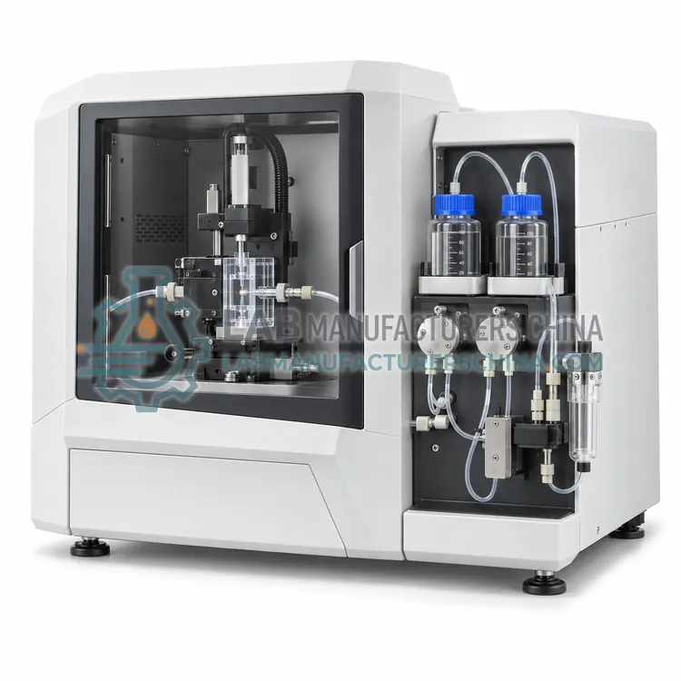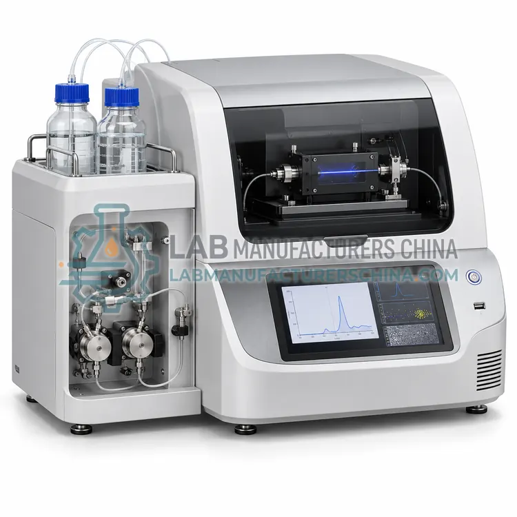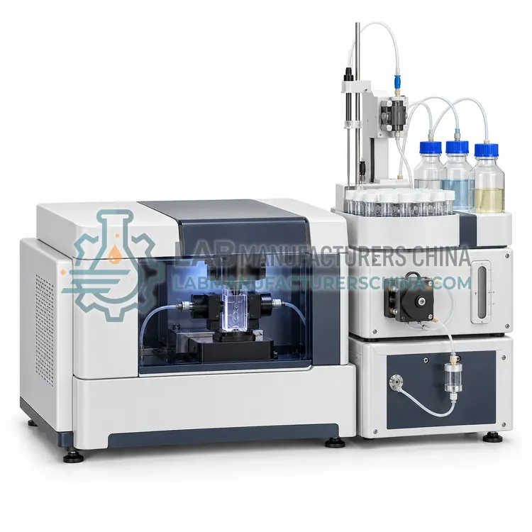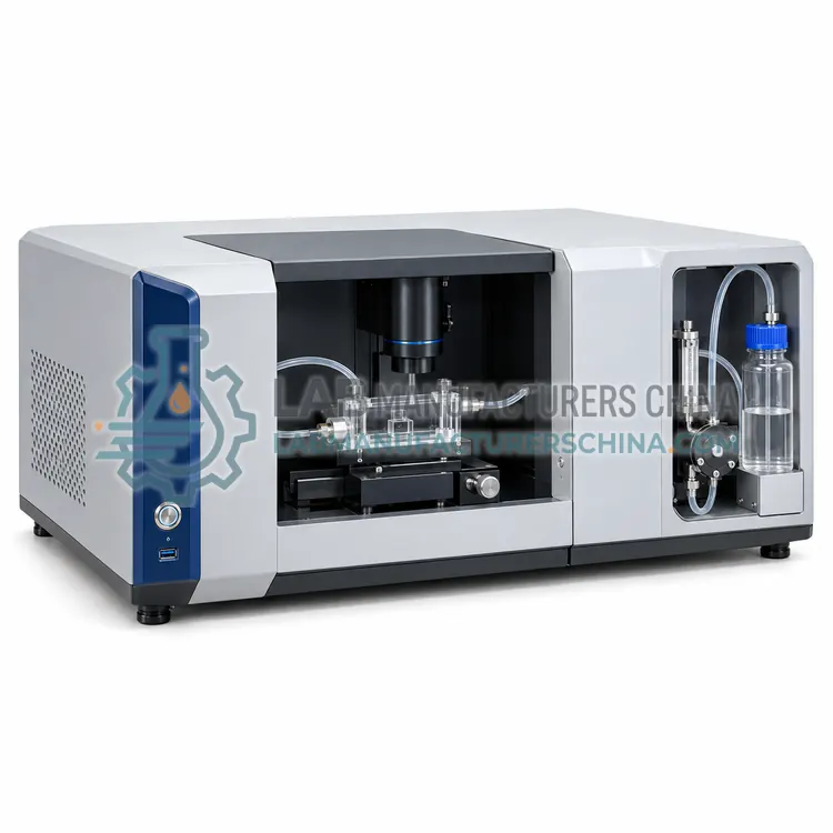
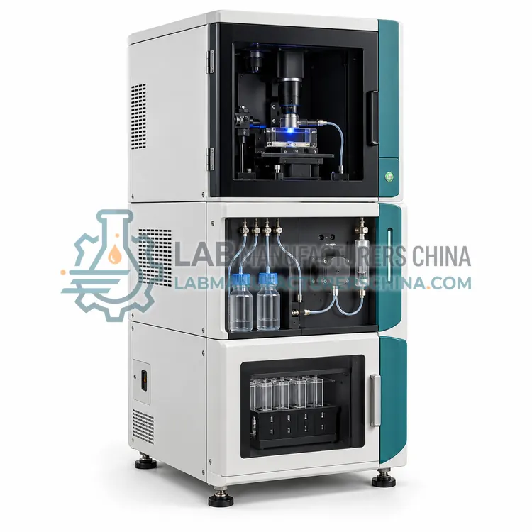
Fluorescence-Mode Nanoparticle Tracking Analyzer
荧光模式纳米颗粒追踪分析仪
- SKU NAA-0002
- Nanoparticle Analyzers
- CE (to be confirmed)
- ISO 9001 manufacturing (to be confirmed)
Price on request — FOB / CIF quoted per order
Prices depend on configuration, quantity and destination port.
Key specifications
| Size range (fluorescence) | ≈ 50–1000 nm (fluorophore dependent) |
|---|---|
| Laser lines | 405, 488, 532, 640 nm (selectable) |
| Emission filters | Long-pass, several cut-on wavelengths |
| Camera | sCMOS, ≥ 30 fps, high sensitivity |
| Concentration range | 10⁷–10⁹ particles/mL |
| Sample volume | 0.3–1 mL |
Request a quote Download brochure · to be provided
By Lab Manufacturers China Technical TeamLast reviewed Standards: ISO 19430, ISO 13322-1, ISO 22412
What is the Fluorescence-Mode Nanoparticle Tracking Analyzer?
A nanoparticle tracking instrument with laser lines and emission filters that distinguishes fluorescently labelled particles from the unlabelled background. It counts and sizes specific subpopulations, such as antibody-labelled vesicles or dye-loaded liposomes, in complex matrices like plasma or cell culture media.
What is the Fluorescence-Mode Nanoparticle Tracking Analyzer?
A nanoparticle tracking instrument with laser lines and emission filters that distinguishes fluorescently labelled particles from the unlabelled background. It counts and sizes specific subpopulations, such as antibody-labelled vesicles or dye-loaded liposomes, in complex matrices like plasma or cell culture media.
Overview
This variant adds switchable long-pass filters and one or more excitation lasers, commonly 405, 488, 532 and 640 nm, to a scattering tracking system. In fluorescence mode only particles emitting within the filter band are tracked, which suppresses contaminants, protein aggregates and unlabelled particles that would swamp the scattering signal. This permits quantification of marker-positive subpopulations, for example CD63-labelled exosomes or fluorescent nanoparticle formulations, in biological fluids. Sensitivity depends on fluorophore brightness and photobleaching; quantum dots and bright organic dyes are favoured. Users must validate labelling efficiency, remove unbound dye, and run fluorescent bead standards. Fluorescence size distributions are typically reported with a detection limit of about 50 to 100 nm, and scattering and fluorescence measurements can be compared on the same sample.
How does it work?
Nanoparticle tracking analysis illuminates a dilute suspension with a laser sheet and records the scattered light of individual particles with a camera; software follows each track and derives a diffusion coefficient, converted to hydrodynamic diameter with the Stokes–Einstein equation, and counts particles per millilitre in a known volume. Resistive pulse sensing passes particles through a nanopore in an electrolyte; each particle transiently reduces ionic current in proportion to its volume, giving size and concentration. Field-flow fractionation separates particles in a thin channel by diffusion under a cross-flow before in-line light scattering detectors measure size and molar mass. Nano flow cytometers use hydrodynamic focusing and sensitive side-scatter and fluorescence detection to phenotype single particles. Sample dilution and calibration with reference beads are essential.
What are the key features?
- Multiple excitation lasers with switchable emission filters
- Fluorescence-only tracking to ignore unlabelled background
- Scattering and fluorescence comparison on the same sample
- Suitable for plasma and cell culture media matrices
- Fluorescent bead calibration routines
- Temperature-controlled sample chamber with flow option
What are the technical specifications?
A typical fluorescence-mode nanoparticle tracking analyzer configuration offers size range (fluorescence) of ≈ 50–1000 nm (fluorophore dependent), laser lines of 405, 488, 532, 640 nm (selectable), emission filters of Long-pass, several cut-on wavelengths, camera of sCMOS, ≥ 30 fps, high sensitivity, concentration range of 10⁷–10⁹ particles/mL and sample volume of 0.3–1 mL. Values are indicative of the product class; the exact figures for each configuration are confirmed on the quotation and the calibration documents supplied with the instrument.
Which applications is it used for?
The Fluorescence-Mode Nanoparticle Tracking Analyzer is used in labelled extracellular vesicle quantification, dye-loaded liposome integrity, fluorescent nanoparticle formulation QC, viral vector particle subsets and immunolabelled biomarker studies. Across the wider Nanoparticle Analyzers class, laboratories also deploy it for liposome and lipid nanoparticle characterisation, extracellular vesicle and exosome research and viral vector and vaccine particle counting.
Which standards and certifications apply?
Relevant standards and methods include ISO 19430, ISO 13322-1, ISO 22412, ISO/TS 21362, ASTM E2834, ISO 21363 and GB/T 19627. Certification candidates for this model are CE and ISO 9001 manufacturing; each is to be confirmed for the specific configuration before shipment, and certificates are supplied only where they have been issued.
Nanoparticle measurements are very sensitive to sample preparation. Use particle-free, filtered diluents and clean glassware, and dilute to the instrument's optimum concentration window. Dust and bubbles produce false counts. Verify size and concentration with certified polystyrene or silica nanosphere standards at start-up and after changes of settings, and report camera level, detection threshold, temperature and number of tracks, because these parameters change results. Keep laser optics, flow cells and nanopores clean and stored as advised. Install in a stable, temperature-controlled room. Regulated work needs IQ/OQ/PQ, locked methods and audit trails.
How do I select the right Fluorescence-Mode Nanoparticle Tracking Analyzer?
Choose fluorescence mode when specific labelled particles must be counted in a complex background, for example exosomes in plasma. Specify laser wavelengths, fluorophores used, sensitivity requirements, sample matrix and whether scattering mode is also needed. Unlabelled, simple suspensions can be measured with the basic scattering variant.
How does it compare with other Nanoparticle Analyzers variants?
Decide what the measurement must deliver: concentration and size distribution of polydisperse samples, fluorescent subpopulation counts, high-resolution separation of mixtures, or single-particle phenotype. Nanoparticle tracking suits 30 to 1000 nm particles at 10⁷ to 10⁹ particles/mL. Resistive pulse sensing gives high-resolution, refractive-index-independent size for conductive dispersions. Field-flow fractionation coupled to multi-angle light scattering resolves mixtures and aggregates. Nano flow cytometry is preferred when labelling and marker analysis are needed. Check detection limit versus your smallest particle, required dynamic range, sample volume, and optical property dependence. Consider whether applicable guidance such as ISO 19430 and regulatory expectations for liposomes or vaccines apply, and who will operate and maintain the system.
Full specifications
| Size range (fluorescence) | ≈ 50–1000 nm (fluorophore dependent) |
|---|---|
| Laser lines | 405, 488, 532, 640 nm (selectable) |
| Emission filters | Long-pass, several cut-on wavelengths |
| Camera | sCMOS, ≥ 30 fps, high sensitivity |
| Concentration range | 10⁷–10⁹ particles/mL |
| Sample volume | 0.3–1 mL |
| Temperature control | 15–55 °C |
| Size accuracy | ± 5% on fluorescent standard |
| Modes | Scatter and fluorescence |
| Data output | Size, concentration, video, track files |
Indicative values for this product class. Final specifications are confirmed on the quotation and in the calibration documents supplied with the instrument.
Applications
- Labelled extracellular vesicle quantification
- Dye-loaded liposome integrity
- Fluorescent nanoparticle formulation QC
- Viral vector particle subsets
- Immunolabelled biomarker studies
Key features
- Multiple excitation lasers with switchable emission filters
- Fluorescence-only tracking to ignore unlabelled background
- Scattering and fluorescence comparison on the same sample
- Suitable for plasma and cell culture media matrices
- Fluorescent bead calibration routines
- Temperature-controlled sample chamber with flow option
Frequently asked questions about Fluorescence-Mode Nanoparticle Tracking Analyzer
Only particles with enough label emit detectable light, so counts reflect the labelled subpopulation and depend on labelling efficiency and photobleaching. Lower counts than scatter mode are expected and useful information.
Remove free dye by size-exclusion chromatography, ultrafiltration or dialysis before analysis, and run a dye-only control. Residual dye can form micelles or aggregates that appear as false particles.
Sequential acquisition with different laser and filter sets is used to measure each channel, because this system images one channel at a time. Co-localisation of markers on the same particle requires a different technique such as nano flow cytometry.
Most cover about 1 to 1000 nm, but each technique has its own window. Tracking analysis typically detects 30 to 1000 nm, resistive pulse sensing 50 nm to several micrometres depending on pore, and field-flow fractionation 1 nm to 1 µm.
By counting particles in a known illuminated or sensed volume, for example particles per millilitre in tracking analysis, or pulse frequency at a known flow rate in resistive pulse sensing. Accuracy depends on dilution, detection threshold and calibration with reference particles.
Tracking follows individual particles and provides number-weighted sizes and concentration, resolving polydispersity better. Dynamic light scattering measures the ensemble signal with intensity weighting, is faster and works at smaller sizes, but does not report concentration.
Related models